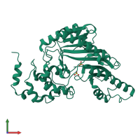
1g0w

X-ray diffraction
2.3Å resolution

CRYSTAL STRUCTURE OF BOVINE RETINAL CREATINE KINASE

Released:
Source organism: Bos taurus
Primary publication:
The three-dimensional structure of cytosolic bovine retinal creatine kinase.
Acta Crystallogr D Biol Crystallogr 57 187-93 (2001)
PMID: 11173463

Function and Biology Details

Structure analysis Details

Assembly composition:
monomeric (preferred)
Assembly name:
PDBe Complex ID:
PDB-CPX-195033 (preferred)
Entry contents:
1 distinct polypeptide molecule
Macromolecule:
Creatine kinase M-type Chain: A
Molecule details ›
Chain: A
Length: 380 amino acids
Theoretical weight: 42.63 KDa
Source organism: Bos taurus
UniProt:
  • Canonical: Q9XSC6 (Residues: 2-381; Coverage: 100%)
Gene name: CKM
Sequence domains:
Structure domains:

Ligands and Environments

1 bound ligand:
No modified residues

Experiments and Validation Details

Entry percentile scores
X-ray source: SRS BEAMLINE PX9.6
Spacegroup: P43212
Unit cell:
a: 95.672Å b: 95.672Å c: 107.913Å
α: 90° β: 90° γ: 90°
R-values:
R R work R free
0.232 0.197 0.232