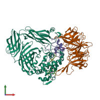
Function and Biology Details
Biochemical function:
Biological process:
- not assigned
Cellular component:
Sequence domains:
- Cytochrome c-like domain superfamily
- Immunoglobulin-like fold
- Immunoglobulin E-set
- Quinohemoprotein amine dehydrogenase alpha subunit, domain 2 superfamily
- Quinohemoprotein amine dehydrogenase, gamma subunit, structural domain
- Quinohemoprotein amine dehydrogenase subunit gamma
- Quinohemoprotein amine dehydrogenase, gamma subunit structural domain superfamily
- Quinohemoprotein amine dehydrogenase, beta subunit
7 more domains
Structure analysis Details
Assemblies composition:
Assembly name:
Quinohemoprotein amine dehydrogenase (preferred)
PDBe Complex ID:
PDB-CPX-140974 (preferred)
Entry contents:
3 distinct polypeptide molecules
Macromolecules (3 distinct):


