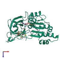
Function and Biology Details
Reaction catalysed:
2 reduced ferredoxin + NADP(+) + H(+) = 2 oxidized ferredoxin + NADPH
Biochemical function:
Biological process:
- not assigned
Cellular component:
- not assigned
Sequence domains:
- Flavoprotein pyridine nucleotide cytochrome reductase
- Ferredoxin-NADP reductase (FNR), nucleotide-binding domain
- Ferredoxin--NADP reductase
- Oxidoreductase FAD/NAD(P)-binding
- FAD-binding domain, ferredoxin reductase-type
- Riboflavin synthase-like beta-barrel
- Ferredoxin--NADP reductase, plant and Cyanobacteria type
Structure domains:
Structure analysis Details
Assembly composition:
monomeric (preferred)
Assembly name:
Ferredoxin--NADP reductase (preferred)
PDBe Complex ID:
PDB-CPX-149470 (preferred)
Entry contents:
1 distinct polypeptide molecule
Macromolecule:


