1s1c

X-ray diffraction
2.6Å resolution

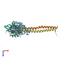
Crystal structure of the complex between the human RhoA and Rho-binding domain of human ROCKI

Released:
Source organism: Homo sapiens

Function and Biology Details

Reactions catalysed:
GTP + H(2)O = GDP + phosphate
ATP + a protein = ADP + a phosphoprotein

Structure analysis Details

Assembly composition:
hetero tetramer (preferred)
PDBe Complex ID:
PDB-CPX-158292 (preferred)
Entry contents:
2 distinct polypeptide molecules
Macromolecules (2 distinct):
Transforming protein RhoA Chains: A, B
Molecule details ›
Chains: A, B
Length: 183 amino acids
Theoretical weight: 20.61 KDa
Source organism: Homo sapiens
Expression system: Escherichia coli
UniProt:
  • Canonical: P61586 (Residues: 1-181; Coverage: 94%)
Gene names: ARH12, ARHA, RHO12, RHOA
Sequence domains: Ras family
Structure domains: P-loop containing nucleotide triphosphate hydrolases
Rho-associated protein kinase 1 Chains: X, Y
Molecule details ›
Chains: X, Y
Length: 71 amino acids
Theoretical weight: 8.45 KDa
Source organism: Homo sapiens
Expression system: Escherichia coli
UniProt:
  • Canonical: Q13464 (Residues: 947-1015; Coverage: 5%)
Gene name: ROCK1
Sequence domains: Rho Binding
Structure domains: Single helix bin

Ligands and Environments

2 bound ligands:
No modified residues

Experiments and Validation Details

Entry percentile scores
X-ray source: ESRF BEAMLINE ID29
Spacegroup: P21
Unit cell:
a: 34.404Å b: 89.537Å c: 98.105Å
α: 90° β: 91.86° γ: 90°
R-values:
R R work R free
0.224 0.219 0.257
Expression system: Escherichia coli