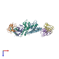
2a0l

X-ray diffraction
3.9Å resolution

Crystal structure of KvAP-33H1 Fv complex

Released:
Source organisms:
Primary publication:
Structure of the KvAP voltage-dependent K+ channel and its dependence on the lipid membrane.
Proc Natl Acad Sci U S A 102 15441-6 (2005)
PMID: 16223877

Function and Biology Details

Structure analysis Details

Assembly composition:
hetero dodecamer (preferred)
PDBe Complex ID:
PDB-CPX-212897 (preferred)
Entry contents:
3 distinct polypeptide molecules
Macromolecules (3 distinct):
Voltage-gated potassium channel Chains: A, B
Molecule details ›
Chains: A, B
Length: 241 amino acids
Theoretical weight: 26.17 KDa
Source organism: Aeropyrum pernix
Expression system: Escherichia coli
UniProt:
  • Canonical: Q9YDF8 (Residues: 20-259; Coverage: 81%)
Gene name: APE_0955
Sequence domains:
Structure domains: Helix Hairpins
33H1 Fv fragment Chains: C, E
Molecule details ›
Chains: C, E
Length: 105 amino acids
Theoretical weight: 11.24 KDa
Source organism: Mus musculus
Structure domains: Immunoglobulins
33H1 Fv fragment Chains: D, F
Molecule details ›
Chains: D, F
Length: 116 amino acids
Theoretical weight: 13.18 KDa
Source organism: Mus musculus
Structure domains: Immunoglobulins

Ligands and Environments

1 bound ligand:
No modified residues

Experiments and Validation Details

Entry percentile scores
X-ray source: NSLS BEAMLINE X25, ALS BEAMLINE 8.2.1, CHESS BEAMLINE A1
Spacegroup: P4222
Unit cell:
a: 136.362Å b: 136.362Å c: 191.812Å
α: 90° β: 90° γ: 90°
R-values:
R R work R free
0.358 0.358 0.392
Expression system: Escherichia coli