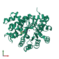
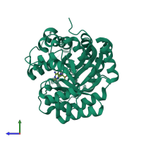
2z7g

X-ray diffraction
2.52Å resolution

Crystal structure of adenosine deaminase ligated with EHNA

Released:
Source organism: Bos taurus
Primary publication:
Conformational change of adenosine deaminase during ligand-exchange in a crystal.
Biochem Biophys Res Commun 373 53-7 (2008)
PMID: 18549808

Function and Biology Details

Reaction catalysed:
2'-deoxyadenosine + H(2)O = 2'-deoxyinosine + NH(3)

Structure analysis Details

Assembly composition:
monomeric (preferred)
Assembly name:
PDBe Complex ID:
PDB-CPX-157515 (preferred)
Entry contents:
1 distinct polypeptide molecule
Macromolecule:
Adenosine deaminase Chain: A
Molecule details ›
Chain: A
Length: 356 amino acids
Theoretical weight: 40.34 KDa
Source organism: Bos taurus
UniProt:
  • Canonical: P56658 (Residues: 2-357; Coverage: 98%)
Gene name: ADA
Sequence domains: Adenosine deaminase
Structure domains: Metal-dependent hydrolases

Ligands and Environments

2 bound ligands:
No modified residues

Experiments and Validation Details

Entry percentile scores
X-ray source: RIGAKU ULTRAX 18
Spacegroup: P43212
Unit cell:
a: 77.1Å b: 77.1Å c: 135.66Å
α: 90° β: 90° γ: 90°
R-values:
R R work R free
0.22 0.207 0.247