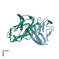
3fc0

X-ray diffraction
1.76Å resolution

1.8 A crystal structure of murine GITR ligand dimer expressed in Drosophila melanogaster S2 cells

Released:
Source organism: Mus musculus
Primary publication:
1.8 A structure of murine GITR ligand dimer expressed in Drosophila melanogaster S2 cells.
Acta Crystallogr D Biol Crystallogr 65 434-9 (2009)
PMID: 19390148

Function and Biology Details

Structure analysis Details

Assembly composition:
homo dimer (preferred)
PDBe Complex ID:
PDB-CPX-182023 (preferred)
Entry contents:
1 distinct polypeptide molecule
Macromolecule:
Tumor necrosis factor ligand superfamily member 18 Chains: A, B
Molecule details ›
Chains: A, B
Length: 139 amino acids
Theoretical weight: 15.9 KDa
Source organism: Mus musculus
Expression system: Drosophila melanogaster
UniProt:
  • Canonical: Q7TS55 (Residues: 46-173; Coverage: 74%)
Gene names: Gitrl, Tnfsf18
Sequence domains: TNF(Tumour Necrosis Factor) family
Structure domains: Jelly Rolls

Ligands and Environments

1 bound ligand:
No modified residues

Experiments and Validation Details

Entry percentile scores
X-ray source: NSLS BEAMLINE X29A
Spacegroup: P212121
Unit cell:
a: 52.609Å b: 69.293Å c: 73.994Å
α: 90° β: 90° γ: 90°
R-values:
R R work R free
0.226 0.224 0.262
Expression system: Drosophila melanogaster