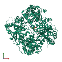
3tuv

X-ray diffraction
2.27Å resolution

Crystal structure of insulysin with bound ATP

Released:
Model geometry
Fit model/data
Source organism: Rattus norvegicus
Primary publication:
Anion activation site of insulin-degrading enzyme.
J Biol Chem 287 48-57 (2012)
PMID: 22049080

Function and Biology Details

Reaction catalysed:
Degradation of insulin, glucagon and other polypeptides. No action on proteins.
Biochemical function:
Cellular component:

Structure analysis Details

Assembly composition:
hetero dimer (preferred)
PDBe Complex ID:
PDB-CPX-152946 (preferred)
Entry contents:
2 distinct polypeptide molecules
Macromolecules (2 distinct):
Insulin-degrading enzyme Chain: A
Molecule details ›
Chain: A
Length: 1019 amino acids
Theoretical weight: 117.86 KDa
Source organism: Rattus norvegicus
Expression system: Spodoptera frugiperda
UniProt:
  • Canonical: P35559 (Residues: 1-1019; Coverage: 100%)
Gene name: Ide
Sequence domains:
Structure domains: Metalloenzyme, LuxS/M16 peptidase-like
Peptide Chain: B
Molecule details ›
Chain: B
Length: 3 amino acids
Theoretical weight: 273 Da
Source organism: Rattus norvegicus
Expression system: Spodoptera frugiperda

Ligands and Environments

1 bound ligand:
No modified residues

Experiments and Validation Details

wwPDB Validation report is not available for this entry.
X-ray source: NSLS BEAMLINE X4A
Spacegroup: C2
Unit cell:
a: 114.984Å b: 70.66Å c: 114.06Å
α: 90° β: 92.63° γ: 90°
R-values:
R R work R free
0.22 0.217 0.268
Expression system: Spodoptera frugiperda