3w1m

X-ray diffraction
1.9Å resolution

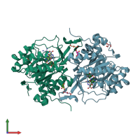
Crystal structure of Trypanosoma cruzi dihydroorotate dehydrogenase in complex with 5-bromoorotate

Released:
Entry authors: Inaoka DK, Iida M, Tabuchi T, Lee N, Matsuoka S, Shiba T, Sakamoto K, Suzuki S, Balogun EO, Nara T, Aoki T, Inoue M, Honma T, Tanaka A

Function and Biology Details

Reaction catalysed:
(S)-dihydroorotate + fumarate = orotate + succinate
Cellular component:

Structure analysis Details

Assembly composition:
homo dimer (preferred)
PDBe Complex ID:
PDB-CPX-175459 (preferred)
Entry contents:
1 distinct polypeptide molecule
Macromolecule:
Dihydroorotate dehydrogenase (fumarate) Chains: A, B
Molecule details ›
Chains: A, B
Length: 313 amino acids
Theoretical weight: 34.07 KDa
Source organism: Trypanosoma cruzi strain CL Brener
Expression system: Escherichia coli
UniProt:
  • Canonical: Q4D3W2 (Residues: 2-314; Coverage: 100%)
Gene names: Tc00.1047053508375.50, pyr4, tcdhod2
Sequence domains: Dihydroorotate dehydrogenase
Structure domains:

Ligands and Environments


Cofactor: Ligand FMN 2 x FMN
3 bound ligands:
No modified residues

Experiments and Validation Details

Entry percentile scores
X-ray source: PHOTON FACTORY BEAMLINE BL-5A
Spacegroup: P212121
Unit cell:
a: 68.341Å b: 71.912Å c: 123.849Å
α: 90° β: 90° γ: 90°
R-values:
R R work R free
0.145 0.143 0.183
Expression system: Escherichia coli