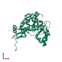
4kie

X-ray diffraction
1.7Å resolution

Crystal structure of the EAL domain of c-di-GMP specific phosphodiesterase YahA

Released:
Model geometry
Fit model/data

Function and Biology Details

Reaction catalysed:
Cyclic di-3',5'-guanylate + H(2)O = 5'-phosphoguanylyl(3'->5')guanosine
Biological process:
  • not assigned
Cellular component:
  • not assigned

Structure analysis Details

Assembly composition:
monomeric (preferred)
PDBe Complex ID:
PDB-CPX-149277 (preferred)
Entry contents:
1 distinct polypeptide molecule
Macromolecule:
Cyclic di-GMP phosphodiesterase PdeL Chain: A
Molecule details ›
Chain: A
Length: 279 amino acids
Theoretical weight: 31.24 KDa
Source organism: Escherichia coli K-12
Expression system: Escherichia coli
UniProt:
  • Canonical: P21514 (Residues: 96-362; Coverage: 74%)
Gene names: JW0307, b0315, pdeL, yahA
Sequence domains: EAL domain
Structure domains: EAL domain

Ligands and Environments

1 bound ligand:
No modified residues

Experiments and Validation Details

wwPDB Validation report is not available for this entry.
X-ray source: SLS BEAMLINE X06DA
Spacegroup: I222
Unit cell:
a: 73.626Å b: 87.818Å c: 94.723Å
α: 90° β: 90° γ: 90°
R-values:
R R work R free
0.166 0.165 0.185
Expression system: Escherichia coli