EMD-0085
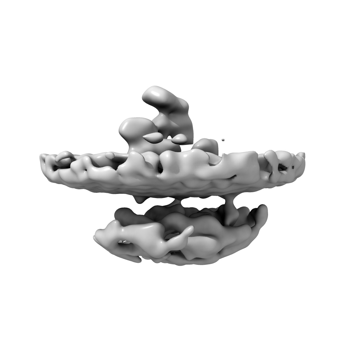
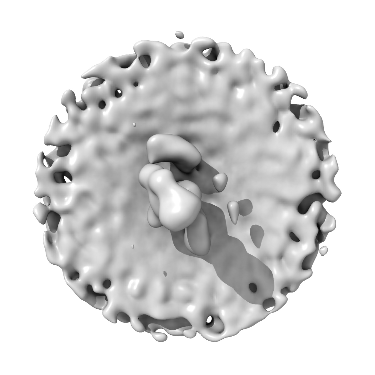
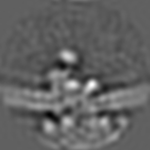
Template-free detection and classification of microsomal membrane bound complexes
EMD-0085
Subtomogram averaging22.0 Å
 Deposition: 27/06/2018
Map released: 02/10/2019
Last modified: 12/02/2020
Sample: Mammalian ribosome bound to the native protein translocon on canine pancreatic ER vesicles
Deposition Authors: Martinez-Sanchez A, Lucic V
Deposition Authors: Martinez-Sanchez A, Lucic V
Template-free detection and classification of membrane-bound complexes in cryo-electron tomograms.
Martinez-Sanchez A  ,
Kochovski Z,
Laugks U ,
Meyer Zum Alten Borgloh J,
Chakraborty S ,
Pfeffer S,
Baumeister W,
Lucic V
,
Kochovski Z,
Laugks U ,
Meyer Zum Alten Borgloh J,
Chakraborty S ,
Pfeffer S,
Baumeister W,
Lucic V
(2020) Nat. Methods , 17 , 209 - 216
 ,
Kochovski Z,
Laugks U ,
Meyer Zum Alten Borgloh J,
Chakraborty S ,
Pfeffer S,
Baumeister W,
Lucic V
,
Kochovski Z,
Laugks U ,
Meyer Zum Alten Borgloh J,
Chakraborty S ,
Pfeffer S,
Baumeister W,
Lucic V
(2020) Nat. Methods , 17 , 209 - 216
Abstract:
With faithful sample preservation and direct imaging of fully hydrated biological material, cryo-electron tomography provides an accurate representation of molecular architecture of cells. However, detection and precise localization of macromolecular complexes within cellular environments is aggravated by the presence of many molecular species and molecular crowding. We developed a template-free image processing procedure for accurate tracing of complex networks of densities in cryo-electron tomograms, a comprehensive and automated detection of heterogeneous membrane-bound complexes and an unsupervised classification (PySeg). Applications to intact cells and isolated endoplasmic reticulum (ER) allowed us to detect and classify small protein complexes. This classification provided sufficiently homogeneous particle sets and initial references to allow subsequent de novo subtomogram averaging. Spatial distribution analysis showed that ER complexes have different localization patterns forming nanodomains. Therefore, this procedure allows a comprehensive detection and structural analysis of complexes in situ.
With faithful sample preservation and direct imaging of fully hydrated biological material, cryo-electron tomography provides an accurate representation of molecular architecture of cells. However, detection and precise localization of macromolecular complexes within cellular environments is aggravated by the presence of many molecular species and molecular crowding. We developed a template-free image processing procedure for accurate tracing of complex networks of densities in cryo-electron tomograms, a comprehensive and automated detection of heterogeneous membrane-bound complexes and an unsupervised classification (PySeg). Applications to intact cells and isolated endoplasmic reticulum (ER) allowed us to detect and classify small protein complexes. This classification provided sufficiently homogeneous particle sets and initial references to allow subsequent de novo subtomogram averaging. Spatial distribution analysis showed that ER complexes have different localization patterns forming nanodomains. Therefore, this procedure allows a comprehensive detection and structural analysis of complexes in situ.