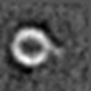
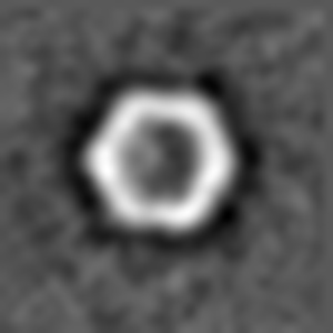
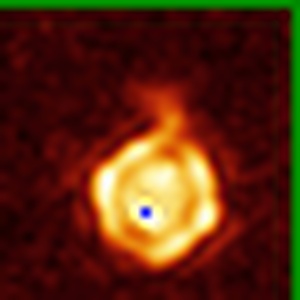
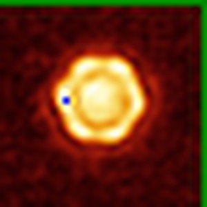
EMD-0621
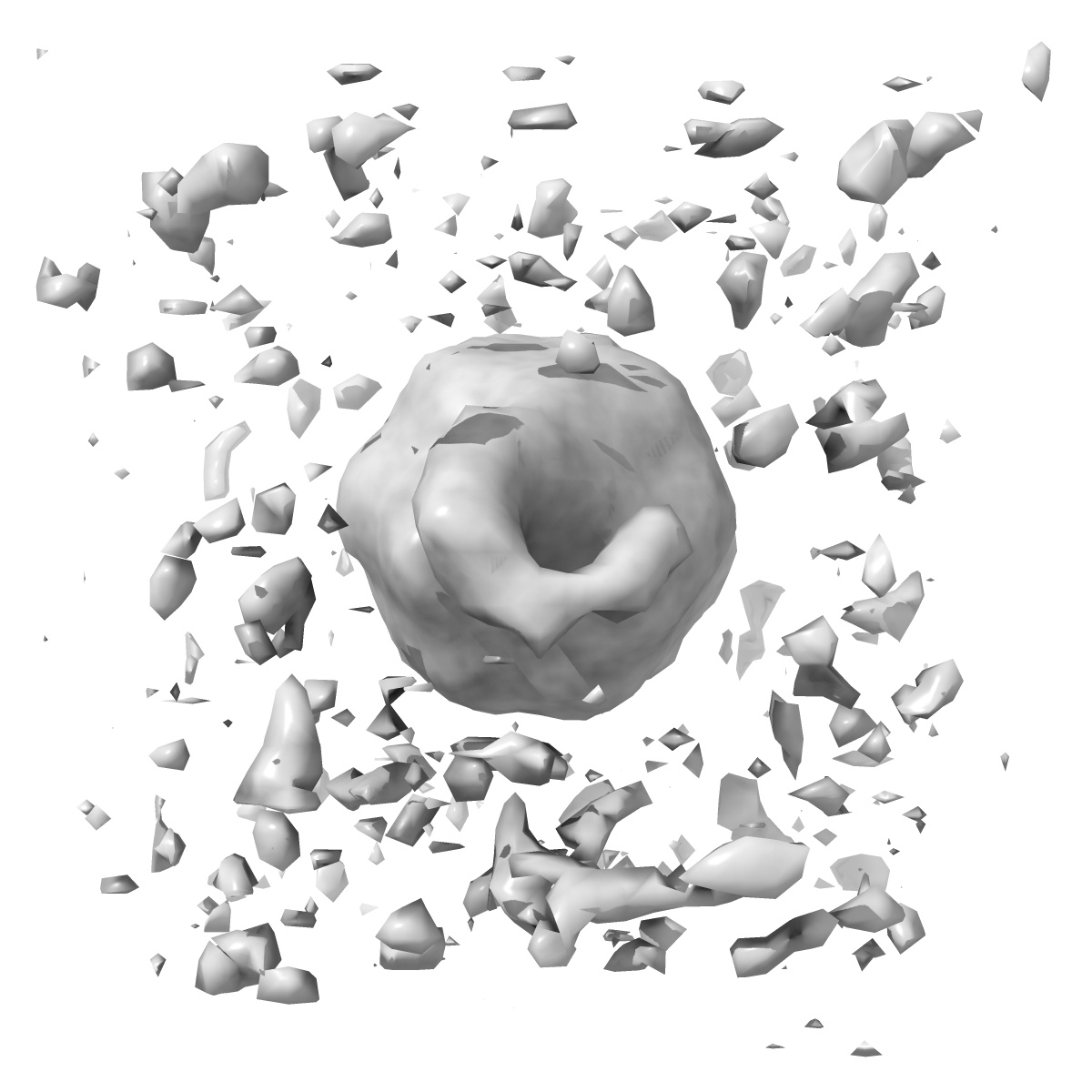
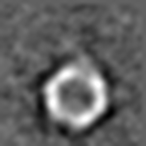
Structure of the AAV2 with its Cell Receptor, AAVR
EMD-0621
Subtomogram averaging20.0 Å
 Deposition: 01/03/2019
Map released: 05/06/2019
Last modified: 05/06/2019
Sample Organism:
Adeno-associated virus
Sample: AAV2 complex with AAVR receptor
Deposition Authors: Hu GQ, Meyer NL, Stagg SM, Chapman MS, Davulcu O, Xie Q, Noble AJ, Yoshioka C, Gingerich D, Trzynka A, David L
Sample: AAV2 complex with AAVR receptor
Deposition Authors: Hu GQ, Meyer NL, Stagg SM, Chapman MS, Davulcu O, Xie Q, Noble AJ, Yoshioka C, Gingerich D, Trzynka A, David L
Structure of the gene therapy vector, adeno-associated virus with its cell receptor, AAVR.
Meyer NL  ,
Hu G,
Davulcu O,
Xie Q,
Noble AJ ,
Yoshioka C ,
Gingerich DS,
Trzynka A,
David L,
Stagg SM ,
Chapman MS
,
Hu G,
Davulcu O,
Xie Q,
Noble AJ ,
Yoshioka C ,
Gingerich DS,
Trzynka A,
David L,
Stagg SM ,
Chapman MS
(2019) eLife , 8
 ,
Hu G,
Davulcu O,
Xie Q,
Noble AJ ,
Yoshioka C ,
Gingerich DS,
Trzynka A,
David L,
Stagg SM ,
Chapman MS
,
Hu G,
Davulcu O,
Xie Q,
Noble AJ ,
Yoshioka C ,
Gingerich DS,
Trzynka A,
David L,
Stagg SM ,
Chapman MS
(2019) eLife , 8
Abstract:
Adeno-associated virus (AAV) vectors are preeminent in emerging clinical gene therapies. Generalizing beyond the most tractable genetic diseases will require modulation of cell specificity and immune neutralization. Interactions of AAV with its cellular receptor, AAVR, are key to understanding cell-entry and trafficking with the rigor needed to engineer tissue-specific vectors. Cryo-electron tomography shows ordered binding of part of the flexible receptor to the viral surface, with distal domains in multiple conformations. Regions of the virus and receptor in close physical proximity can be identified by cross-linking/mass spectrometry. Cryo-electron microscopy with a two-domain receptor fragment reveals the interactions at 2.4 Å resolution. AAVR binds between AAV's spikes on a plateau that is conserved, except in one clade whose structure is AAVR-incompatible. AAVR's footprint overlaps the epitopes of several neutralizing antibodies, prompting a re-evaluation of neutralization mechanisms. The structure provides a roadmap for experimental probing and manipulation of viral-receptor interactions.
Adeno-associated virus (AAV) vectors are preeminent in emerging clinical gene therapies. Generalizing beyond the most tractable genetic diseases will require modulation of cell specificity and immune neutralization. Interactions of AAV with its cellular receptor, AAVR, are key to understanding cell-entry and trafficking with the rigor needed to engineer tissue-specific vectors. Cryo-electron tomography shows ordered binding of part of the flexible receptor to the viral surface, with distal domains in multiple conformations. Regions of the virus and receptor in close physical proximity can be identified by cross-linking/mass spectrometry. Cryo-electron microscopy with a two-domain receptor fragment reveals the interactions at 2.4 Å resolution. AAVR binds between AAV's spikes on a plateau that is conserved, except in one clade whose structure is AAVR-incompatible. AAVR's footprint overlaps the epitopes of several neutralizing antibodies, prompting a re-evaluation of neutralization mechanisms. The structure provides a roadmap for experimental probing and manipulation of viral-receptor interactions.