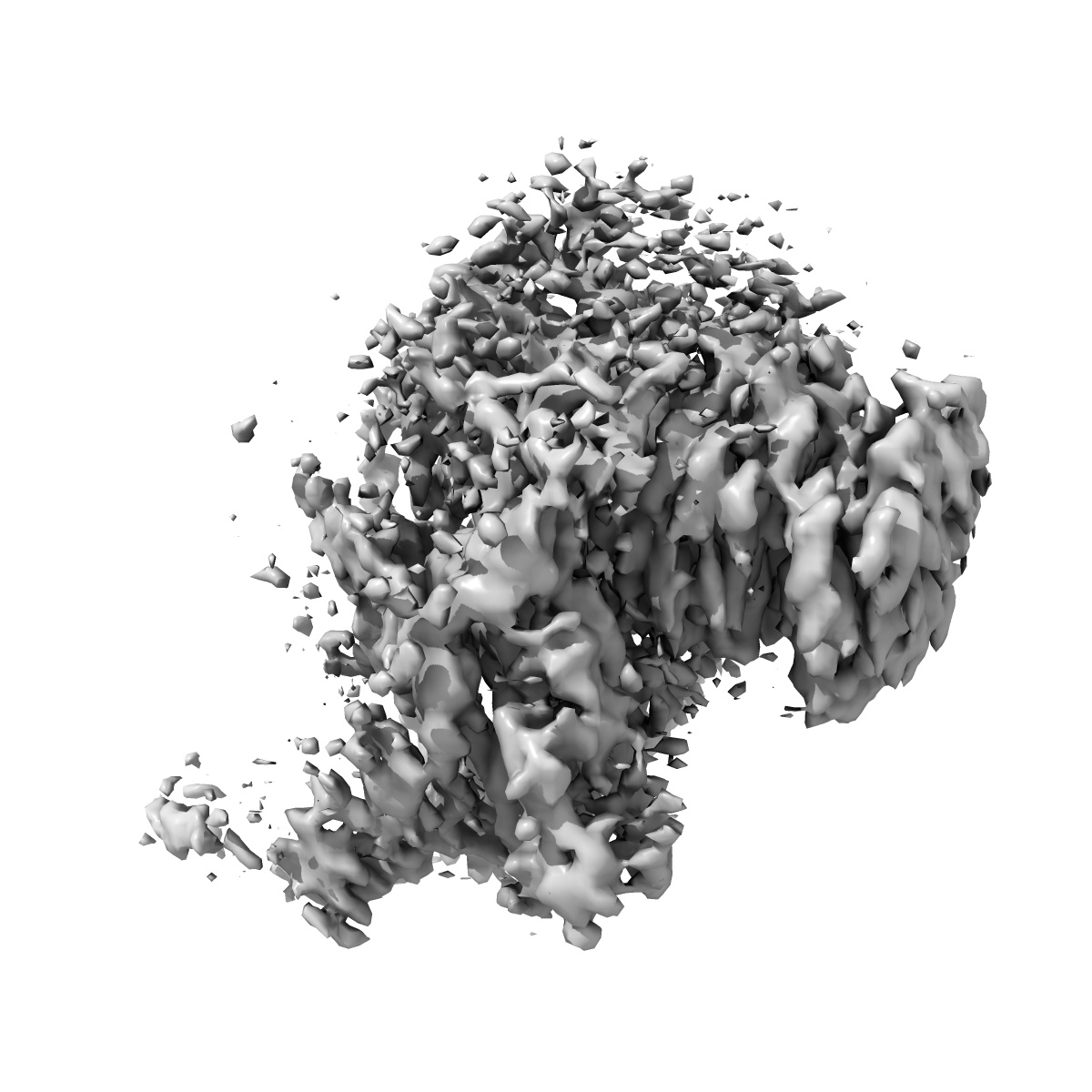
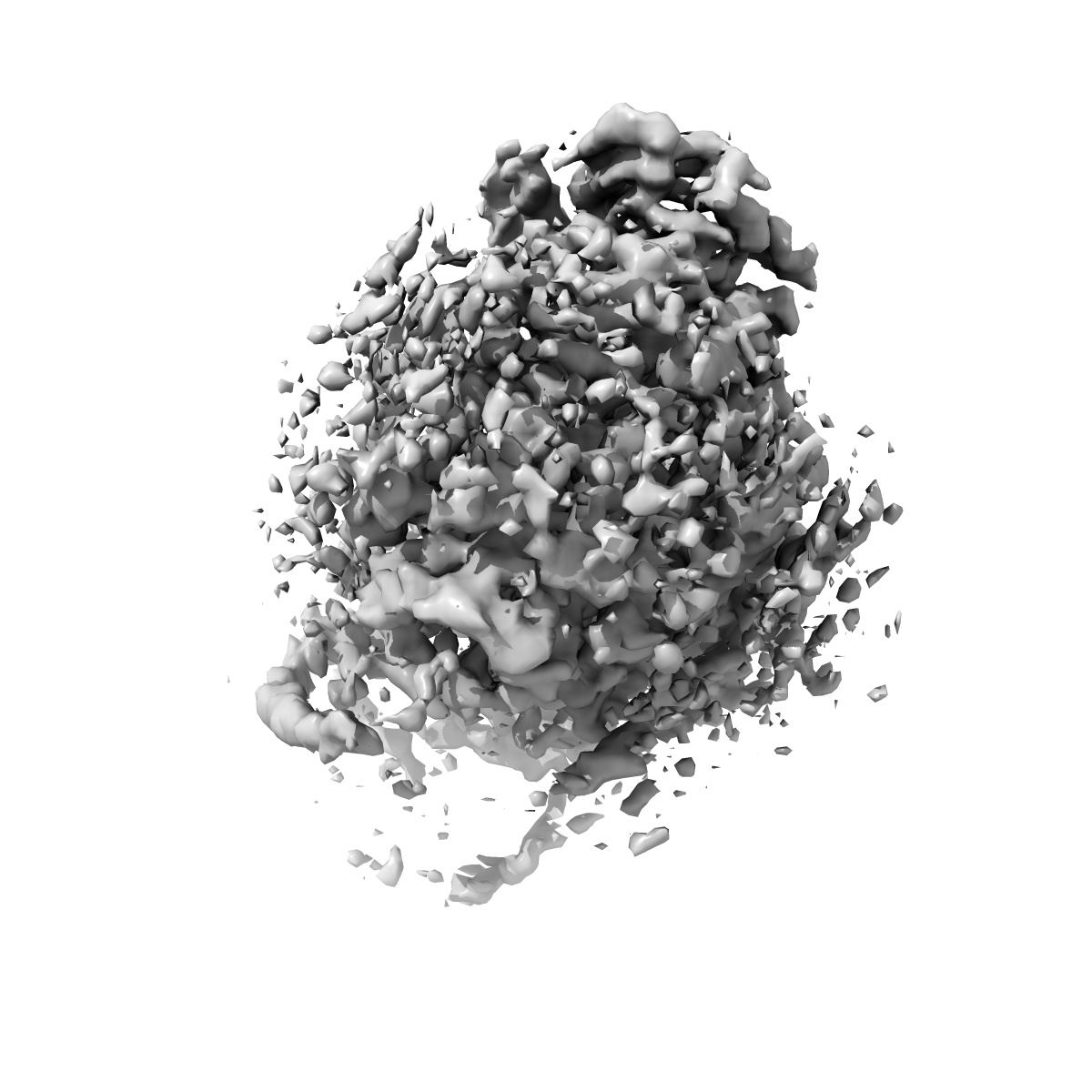
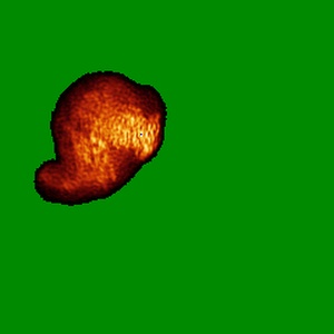
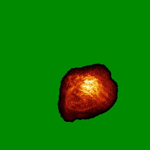
EMD-0670
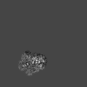
Cryo-EM structure of the mammalian DP-state ATP synthase FO section
EMD-0670
Single-particle4.35 Å
 Deposition: 10/01/2019
Map released: 26/06/2019
Last modified: 26/06/2019
Sample Organism:
Sus
Sample: Cryo-EM structure of the mammalian DP-state ATP synthase FO section
Fitted models: 6j5a (Avg. Q-score: 0.238)
Raw data: EMPIAR-10283
Deposition Authors: Gu JK, Zhang LX, Yi JB, Yang MJ
Sample: Cryo-EM structure of the mammalian DP-state ATP synthase FO section
Fitted models: 6j5a (Avg. Q-score: 0.238)
Raw data: EMPIAR-10283
Deposition Authors: Gu JK, Zhang LX, Yi JB, Yang MJ
Cryo-EM structure of the mammalian ATP synthase tetramer bound with inhibitory protein IF1.
Gu J  ,
Zhang L ,
Zong S ,
Guo R ,
Liu T ,
Yi J ,
Wang P,
Zhuo W ,
Yang M
,
Zhang L ,
Zong S ,
Guo R ,
Liu T ,
Yi J ,
Wang P,
Zhuo W ,
Yang M
(2019) Science , 364 , 1068 - 1075
 ,
Zhang L ,
Zong S ,
Guo R ,
Liu T ,
Yi J ,
Wang P,
Zhuo W ,
Yang M
,
Zhang L ,
Zong S ,
Guo R ,
Liu T ,
Yi J ,
Wang P,
Zhuo W ,
Yang M
(2019) Science , 364 , 1068 - 1075
Abstract:
The mitochondrial adenosine triphosphate (ATP) synthase produces most of the ATP required by mammalian cells. We isolated porcine tetrameric ATP synthase and solved its structure at 6.2-angstrom resolution using a single-particle cryo-electron microscopy method. Two classical V-shaped ATP synthase dimers lie antiparallel to each other to form an H-shaped ATP synthase tetramer, as viewed from the matrix. ATP synthase inhibitory factor subunit 1 (IF1) is a well-known in vivo inhibitor of mammalian ATP synthase at low pH. Two IF1 dimers link two ATP synthase dimers, which is consistent with the ATP synthase tetramer adopting an inhibited state. Within the tetramer, we refined structures of intact ATP synthase in two different rotational conformations at 3.34- and 3.45-Å resolution.
The mitochondrial adenosine triphosphate (ATP) synthase produces most of the ATP required by mammalian cells. We isolated porcine tetrameric ATP synthase and solved its structure at 6.2-angstrom resolution using a single-particle cryo-electron microscopy method. Two classical V-shaped ATP synthase dimers lie antiparallel to each other to form an H-shaped ATP synthase tetramer, as viewed from the matrix. ATP synthase inhibitory factor subunit 1 (IF1) is a well-known in vivo inhibitor of mammalian ATP synthase at low pH. Two IF1 dimers link two ATP synthase dimers, which is consistent with the ATP synthase tetramer adopting an inhibited state. Within the tetramer, we refined structures of intact ATP synthase in two different rotational conformations at 3.34- and 3.45-Å resolution.