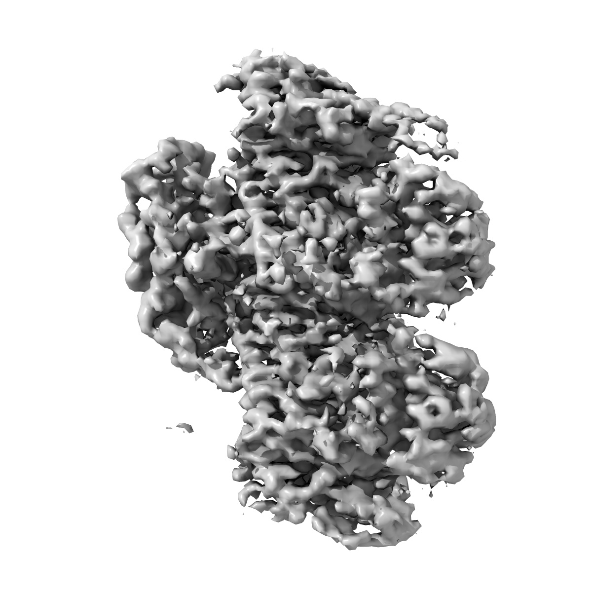
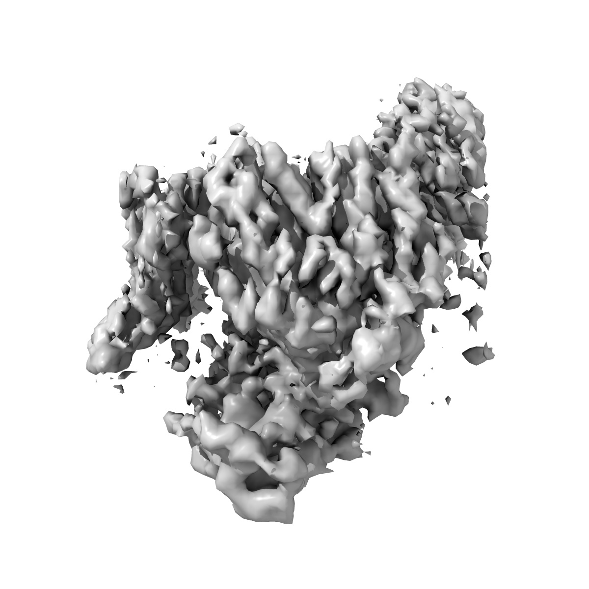
EMD-10131
MKLP2-decocrated 13 protofilament microtubule (ADP.Al.Fx state) processed with MiRP (Microtubule Relion-based Pipeline)
EMD-10131
Single-particle4.2 Å
 Deposition: 18/07/2019
Map released: 23/10/2019
Last modified: 25/11/2020
Sample Organism:
Bos taurus,
Mus musculus
Sample: Complex of 13pf microtubule with bound MKLP2 motor domain in the presence of ADP.AlFx
Raw data: EMPIAR-10796
Deposition Authors: Cook AC, Manka SW, Wang S, Moores CA, Atherton J
Sample: Complex of 13pf microtubule with bound MKLP2 motor domain in the presence of ADP.AlFx
Raw data: EMPIAR-10796
Deposition Authors: Cook AC, Manka SW, Wang S, Moores CA, Atherton J
A microtubule RELION-based pipeline for cryo-EM image processing.

Abstract:
Microtubules are polar filaments built from αβ-tubulin heterodimers that exhibit a range of architectures in vitro and in vivo. Tubulin heterodimers are arranged helically in the microtubule wall but many physiologically relevant architectures exhibit a break in helical symmetry known as the seam. Noisy 2D cryo-electron microscopy projection images of pseudo-helical microtubules therefore depict distinct but highly similar views owing to the high structural similarity of α- and β-tubulin. The determination of the αβ-tubulin register and seam location during image processing is essential for alignment accuracy that enables determination of biologically relevant structures. Here we present a pipeline designed for image processing and high-resolution reconstruction of cryo-electron microscopy microtubule datasets, based in the popular and user-friendly RELION image-processing package, Microtubule RELION-based Pipeline (MiRP). The pipeline uses a combination of supervised classification and prior knowledge about geometric lattice constraints in microtubules to accurately determine microtubule architecture and seam location. The presented method is fast and semi-automated, producing near-atomic resolution reconstructions with test datasets that contain a range of microtubule architectures and binding proteins.
Microtubules are polar filaments built from αβ-tubulin heterodimers that exhibit a range of architectures in vitro and in vivo. Tubulin heterodimers are arranged helically in the microtubule wall but many physiologically relevant architectures exhibit a break in helical symmetry known as the seam. Noisy 2D cryo-electron microscopy projection images of pseudo-helical microtubules therefore depict distinct but highly similar views owing to the high structural similarity of α- and β-tubulin. The determination of the αβ-tubulin register and seam location during image processing is essential for alignment accuracy that enables determination of biologically relevant structures. Here we present a pipeline designed for image processing and high-resolution reconstruction of cryo-electron microscopy microtubule datasets, based in the popular and user-friendly RELION image-processing package, Microtubule RELION-based Pipeline (MiRP). The pipeline uses a combination of supervised classification and prior knowledge about geometric lattice constraints in microtubules to accurately determine microtubule architecture and seam location. The presented method is fast and semi-automated, producing near-atomic resolution reconstructions with test datasets that contain a range of microtubule architectures and binding proteins.