EMD-1040
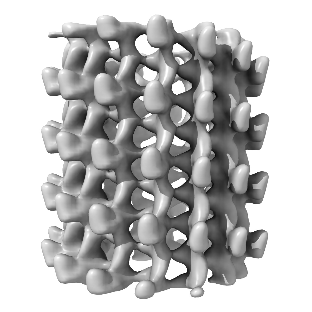
Nucleotide-induced conformations in the neck region of dimeric kinesin.
EMD-1040
Helical reconstruction25.0 Å
 Deposition: 28/02/2003
Map released: 28/03/2003
Last modified: 13/04/2016
Sample Organism:
Rattus norvegicus
Sample: rat kinesin monomer complexed to microtubules in the presence of AMP-PNP
Deposition Authors: Skiniotis G
Sample: rat kinesin monomer complexed to microtubules in the presence of AMP-PNP
Deposition Authors: Skiniotis G
Nucleotide-induced conformations in the neck region of dimeric kinesin.
Skiniotis G  ,
Surrey T ,
Altmann S,
Gross H,
Song YH,
Mandelkow E,
Hoenger A
,
Surrey T ,
Altmann S,
Gross H,
Song YH,
Mandelkow E,
Hoenger A
(2003) Embo J. , 22 , 1518 - 1528
 ,
Surrey T ,
Altmann S,
Gross H,
Song YH,
Mandelkow E,
Hoenger A
,
Surrey T ,
Altmann S,
Gross H,
Song YH,
Mandelkow E,
Hoenger A
(2003) Embo J. , 22 , 1518 - 1528
Abstract:
The neck region of kinesin constitutes a key component in the enzyme's walking mechanism. Here we applied cryoelectron microscopy and image reconstruction to investigate the location of the kinesin neck in dimeric and monomeric constructs complexed to microtubules. To this end we enhanced the visibility of this region by engineering an SH3 domain into the transition between neck linker and neck coiled coil. The resulting chimeric kinesin constructs remained functional as verified by physiology assays. In the presence of AMP-PNP the SH3 domains allowed us to identify the position of the neck in a well defined conformation and revealed its high flexibility in the absence of nucleotide. We show here the double-headed binding of dimeric kinesin along the same protofilament, which is characterized by the opposite directionality of neck linkers. In this configuration the neck coiled coil appears fully zipped. The position of the neck region in dimeric constructs is not affected by the presence of the tubulin C-termini as confirmed by subtilisin treatment of microtubules prior to motor decoration.
The neck region of kinesin constitutes a key component in the enzyme's walking mechanism. Here we applied cryoelectron microscopy and image reconstruction to investigate the location of the kinesin neck in dimeric and monomeric constructs complexed to microtubules. To this end we enhanced the visibility of this region by engineering an SH3 domain into the transition between neck linker and neck coiled coil. The resulting chimeric kinesin constructs remained functional as verified by physiology assays. In the presence of AMP-PNP the SH3 domains allowed us to identify the position of the neck in a well defined conformation and revealed its high flexibility in the absence of nucleotide. We show here the double-headed binding of dimeric kinesin along the same protofilament, which is characterized by the opposite directionality of neck linkers. In this configuration the neck coiled coil appears fully zipped. The position of the neck region in dimeric constructs is not affected by the presence of the tubulin C-termini as confirmed by subtilisin treatment of microtubules prior to motor decoration.