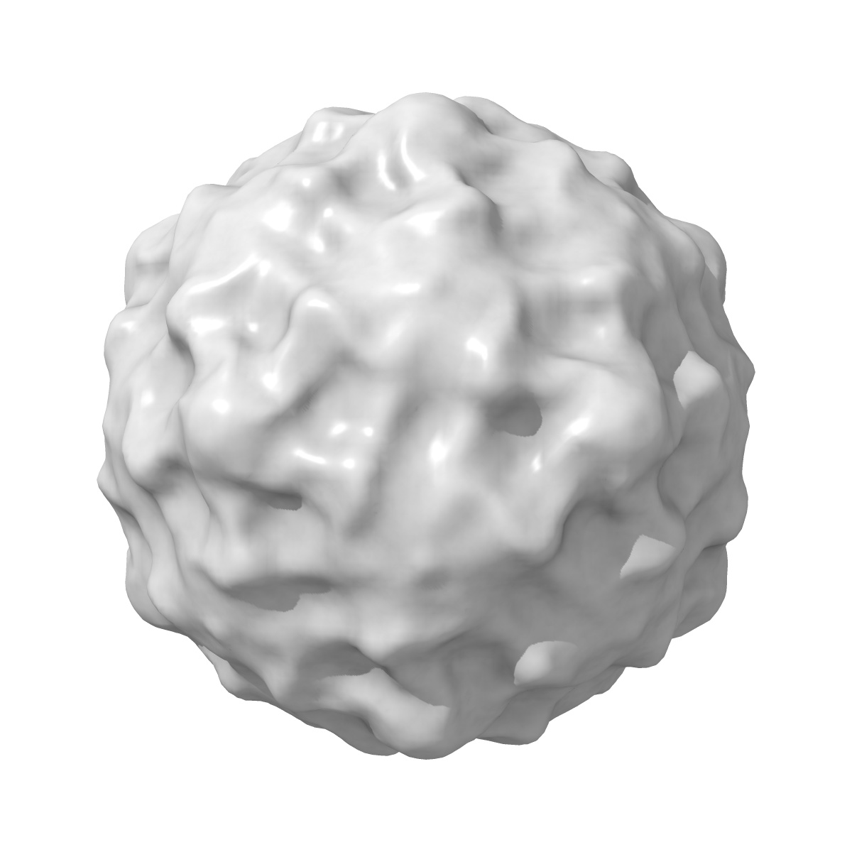
EMD-1057
The structure of echovirus type 12 bound to a two-domain fragment of its cellular attachment protein decay-accelerating factor (CD 55).
EMD-1057
Single-particle16.0 Å
 Deposition: 08/10/2003
Map released: 25/11/2003
Last modified: 26/05/2011
Sample Organism:
Human echovirus 12,
Homo sapiens
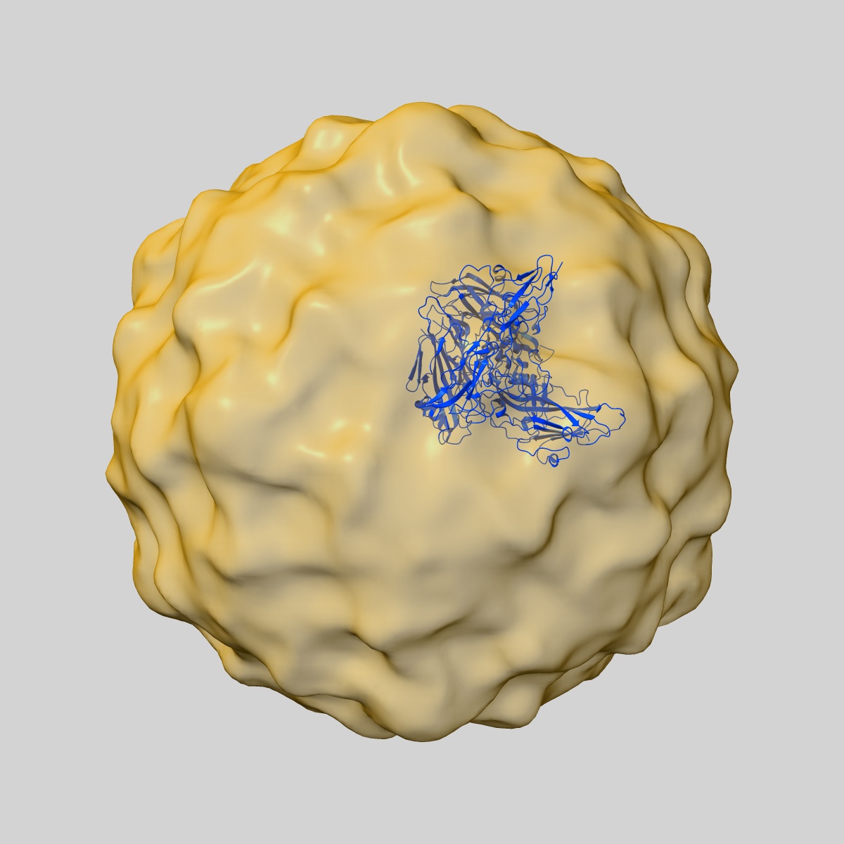
Sample: Echovirus type 12 bound to decay accelerating factor domains 3 and 4
Fitted models: 1upn (Avg. Q-score: 0.032)
Deposition Authors: Bhella D ,
Goodfellow IG ,
Roversi P ,
Pettigrew D,
Chaudhry Y,
Evans DJ ,
Lea SM
,
Goodfellow IG ,
Roversi P ,
Pettigrew D,
Chaudhry Y,
Evans DJ ,
Lea SM
Sample: Echovirus type 12 bound to decay accelerating factor domains 3 and 4
Fitted models: 1upn (Avg. Q-score: 0.032)
Deposition Authors: Bhella D
 ,
Goodfellow IG ,
Roversi P ,
Pettigrew D,
Chaudhry Y,
Evans DJ ,
Lea SM
,
Goodfellow IG ,
Roversi P ,
Pettigrew D,
Chaudhry Y,
Evans DJ ,
Lea SM
The structure of echovirus type 12 bound to a two-domain fragment of its cellular attachment protein decay-accelerating factor (CD 55).
Bhella D ,
Goodfellow IG ,
Roversi P ,
Pettigrew D,
Chaudhry Y,
Evans DJ ,
Lea SM
(2004) J. Biol. Chem. , 279 , 8325 - 8332
,
Goodfellow IG ,
Roversi P ,
Pettigrew D,
Chaudhry Y,
Evans DJ ,
Lea SM
(2004) J. Biol. Chem. , 279 , 8325 - 8332
Abstract:
Echovirus type 12 (EV12), an Enterovirus of the Picornaviridae family, uses the complement regulator decay-accelerating factor (DAF, CD55) as a cellular receptor. We have calculated a three-dimensional reconstruction of EV12 bound to a fragment of DAF consisting of short consensus repeat domains 3 and 4 from cryo-negative stain electron microscopy data (EMD code 1057). This shows that, as for an earlier reconstruction of the related echovirus type 7 bound to DAF, attachment is not within the viral canyon but occurs close to the 2-fold symmetry axes. Despite this general similarity our reconstruction reveals a receptor interaction that is quite different from that observed for EV7. Fitting of the crystallographic co-ordinates for DAF(34) and EV11 into the reconstruction shows a close agreement between the crystal structure of the receptor fragment and the density for the virus-bound receptor, allowing unambiguous positioning of the receptor with respect to the virion (PDB code 1UPN). Our finding that the mode of virus-receptor interaction in EV12 is distinct from that seen for EV7 raises interesting questions regarding the evolution and biological significance of the DAF binding phenotype in these viruses.
Echovirus type 12 (EV12), an Enterovirus of the Picornaviridae family, uses the complement regulator decay-accelerating factor (DAF, CD55) as a cellular receptor. We have calculated a three-dimensional reconstruction of EV12 bound to a fragment of DAF consisting of short consensus repeat domains 3 and 4 from cryo-negative stain electron microscopy data (EMD code 1057). This shows that, as for an earlier reconstruction of the related echovirus type 7 bound to DAF, attachment is not within the viral canyon but occurs close to the 2-fold symmetry axes. Despite this general similarity our reconstruction reveals a receptor interaction that is quite different from that observed for EV7. Fitting of the crystallographic co-ordinates for DAF(34) and EV11 into the reconstruction shows a close agreement between the crystal structure of the receptor fragment and the density for the virus-bound receptor, allowing unambiguous positioning of the receptor with respect to the virion (PDB code 1UPN). Our finding that the mode of virus-receptor interaction in EV12 is distinct from that seen for EV7 raises interesting questions regarding the evolution and biological significance of the DAF binding phenotype in these viruses.