EMD-10752
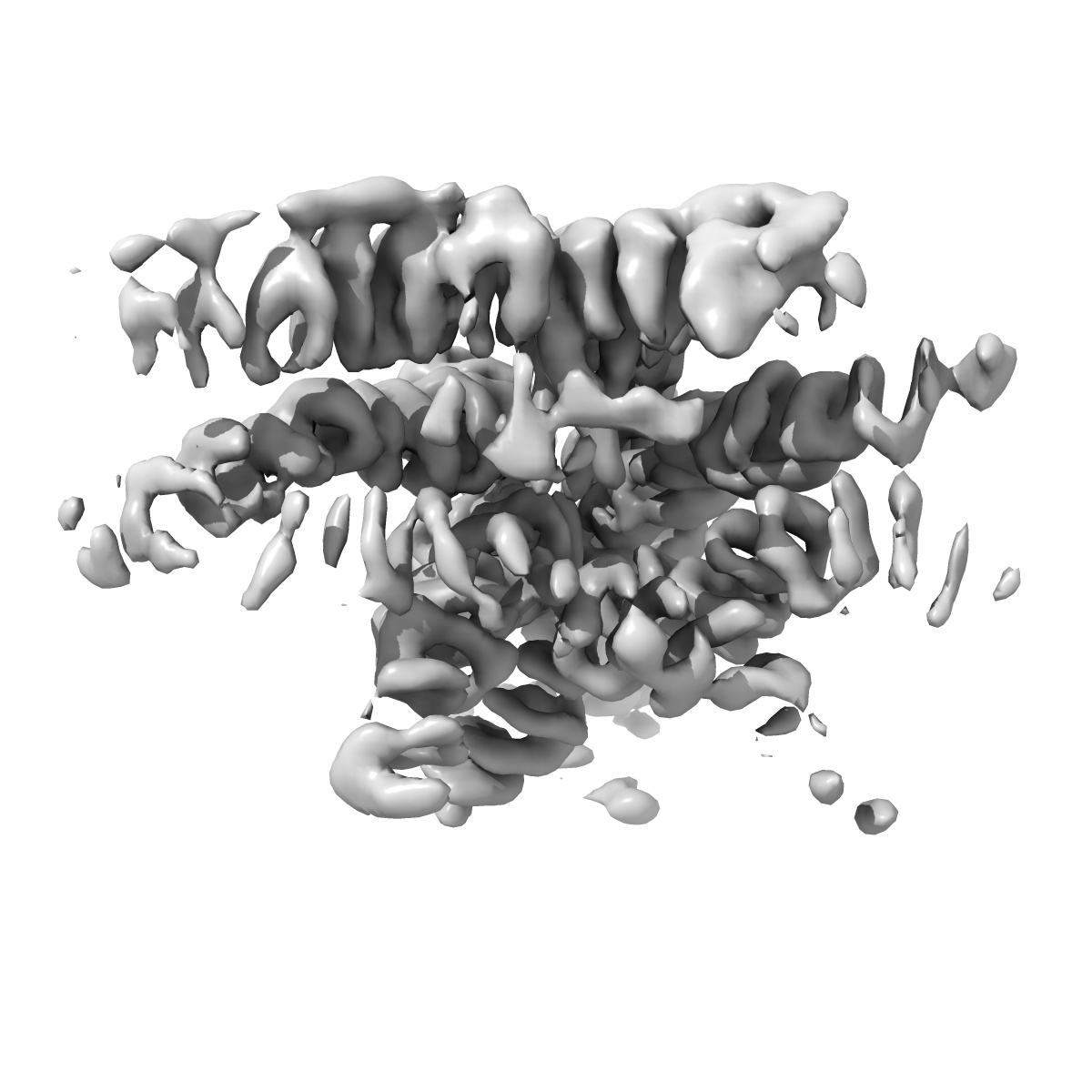
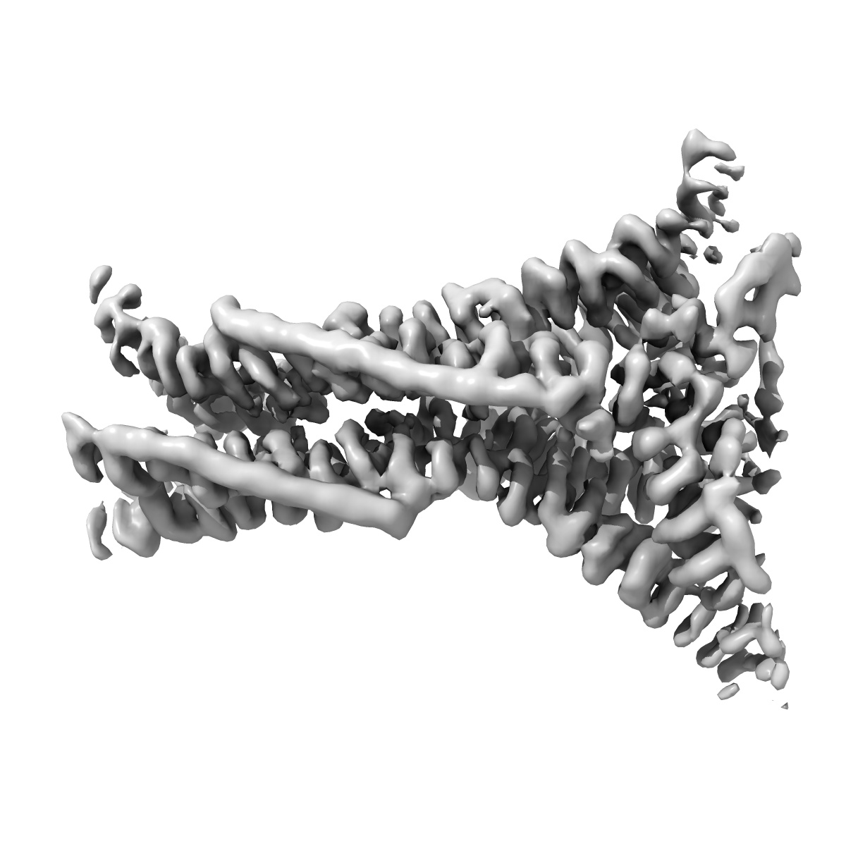
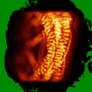
Hexagon bounding clathrin leg in clathrin coats assembled on a membrane
EMD-10752
Subtomogram averaging7.7 Å
 Deposition: 12/03/2020
Map released: 29/07/2020
Last modified: 14/04/2021
Sample Organism:
Sus scrofa
Sample: Clathrin/AP2 coat assembled on membrane.
Deposition Authors: Kovtun O, Kane Dickson V, Kelly BT, Owen D, Briggs JAG
Sample: Clathrin/AP2 coat assembled on membrane.
Deposition Authors: Kovtun O, Kane Dickson V, Kelly BT, Owen D, Briggs JAG
Architecture of the AP2/clathrin coat on the membranes of clathrin-coated vesicles.

Abstract:
Clathrin-mediated endocytosis (CME) is crucial for modulating the protein composition of a cell's plasma membrane. Clathrin forms a cage-like, polyhedral outer scaffold around a vesicle, to which cargo-selecting clathrin adaptors are attached. Adaptor protein complex (AP2) is the key adaptor in CME. Crystallography has shown AP2 to adopt a range of conformations. Here, we used cryo-electron microscopy, tomography, and subtomogram averaging to determine structures, interactions, and arrangements of clathrin and AP2 at the key steps of coat assembly, from AP2 in solution to membrane-assembled clathrin-coated vesicles (CCVs). AP2 binds cargo and PtdIns(4,5)P 2 (phosphatidylinositol 4,5-bisphosphate)-containing membranes via multiple interfaces, undergoing conformational rearrangement from its cytosolic state. The binding mode of AP2 β2 appendage into the clathrin lattice in CCVs and buds implies how the adaptor structurally modulates coat curvature and coat disassembly.
Clathrin-mediated endocytosis (CME) is crucial for modulating the protein composition of a cell's plasma membrane. Clathrin forms a cage-like, polyhedral outer scaffold around a vesicle, to which cargo-selecting clathrin adaptors are attached. Adaptor protein complex (AP2) is the key adaptor in CME. Crystallography has shown AP2 to adopt a range of conformations. Here, we used cryo-electron microscopy, tomography, and subtomogram averaging to determine structures, interactions, and arrangements of clathrin and AP2 at the key steps of coat assembly, from AP2 in solution to membrane-assembled clathrin-coated vesicles (CCVs). AP2 binds cargo and PtdIns(4,5)P 2 (phosphatidylinositol 4,5-bisphosphate)-containing membranes via multiple interfaces, undergoing conformational rearrangement from its cytosolic state. The binding mode of AP2 β2 appendage into the clathrin lattice in CCVs and buds implies how the adaptor structurally modulates coat curvature and coat disassembly.