EMD-1083
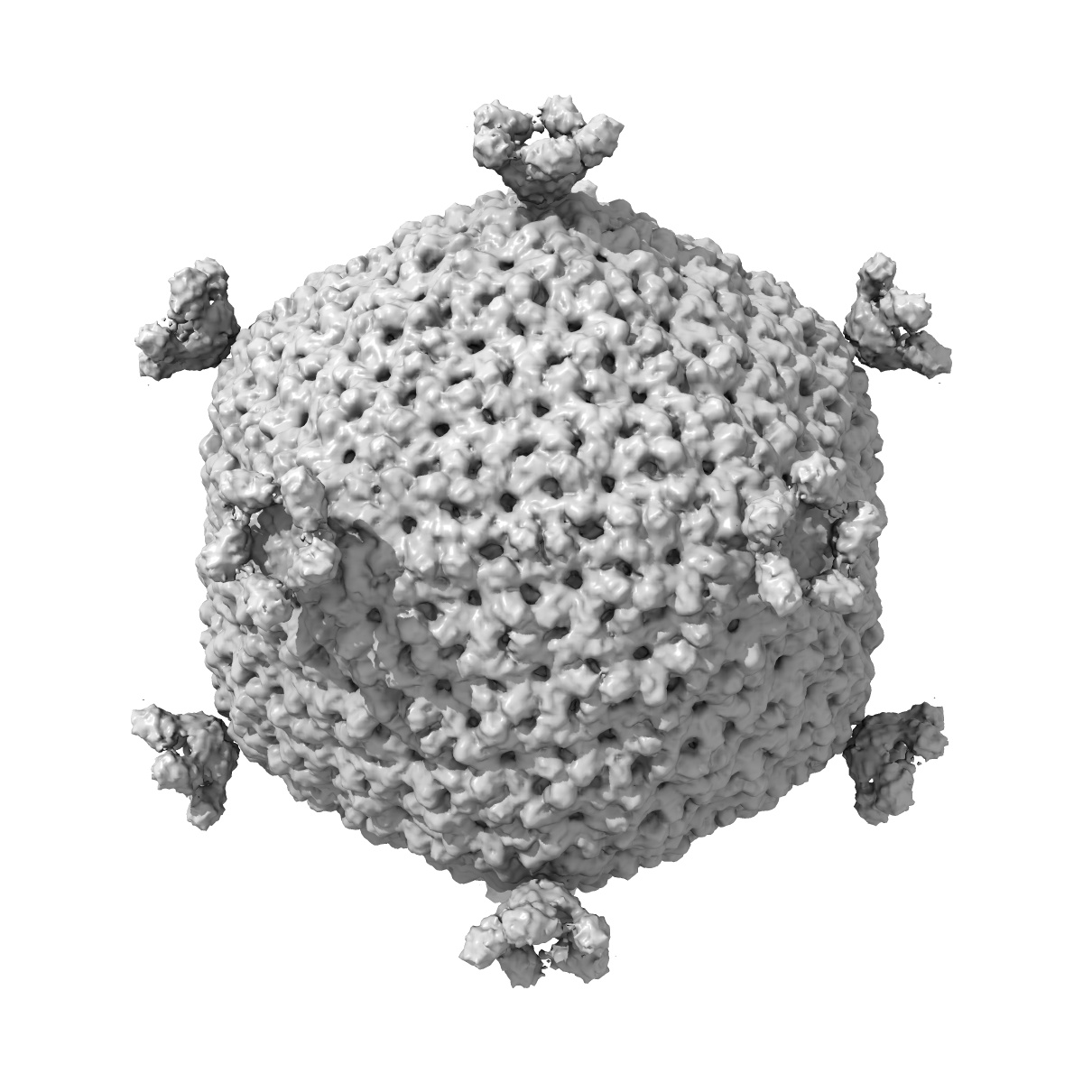
The PM2 virion has a novel organization with an internal membrane and pentameric receptor binding spikes.
EMD-1083
Single-particle12.3 Å
 Deposition: 18/05/2004
Map released: 25/05/2005
Last modified: 17/10/2012
Sample Organism:
Pseudoalteromonas phage PM2
Sample: PM2 virion
Deposition Authors: Huiskonen JT ,
Kivela HM,
Bamford DH ,
Butcher SJ
,
Kivela HM,
Bamford DH ,
Butcher SJ
Sample: PM2 virion
Deposition Authors: Huiskonen JT
 ,
Kivela HM,
Bamford DH ,
Butcher SJ
,
Kivela HM,
Bamford DH ,
Butcher SJ
The PM2 virion has a novel organization with an internal membrane and pentameric receptor binding spikes.
Abstract:
Biological membranes are notoriously resistant to structural analysis. Excellent candidates to tackle this problem in situ are membrane-containing viruses where the membrane is constrained by an icosahedral capsid. Cryo-EM and image reconstruction of bacteriophage PM2 revealed a membrane bilayer following the internal surface of the capsid. The viral genome closely interacts with the inner leaflet. The capsid, at a resolution of 8.4 A, reveals 200 trimeric capsomers with a pseudo T = 21 dextro organization. Pentameric receptor-binding spikes protrude from the surface. It is evident from the structure that the PM2 membrane has at least two important roles in the life cycle. First, it acts as a scaffold to nucleate capsid assembly. Second, after host recognition, it fuses with the host outer membrane to promote genome entry. The structure also sheds light on how the viral supercoiled circular double-stranded DNA genome might be packaged and released.
Biological membranes are notoriously resistant to structural analysis. Excellent candidates to tackle this problem in situ are membrane-containing viruses where the membrane is constrained by an icosahedral capsid. Cryo-EM and image reconstruction of bacteriophage PM2 revealed a membrane bilayer following the internal surface of the capsid. The viral genome closely interacts with the inner leaflet. The capsid, at a resolution of 8.4 A, reveals 200 trimeric capsomers with a pseudo T = 21 dextro organization. Pentameric receptor-binding spikes protrude from the surface. It is evident from the structure that the PM2 membrane has at least two important roles in the life cycle. First, it acts as a scaffold to nucleate capsid assembly. Second, after host recognition, it fuses with the host outer membrane to promote genome entry. The structure also sheds light on how the viral supercoiled circular double-stranded DNA genome might be packaged and released.