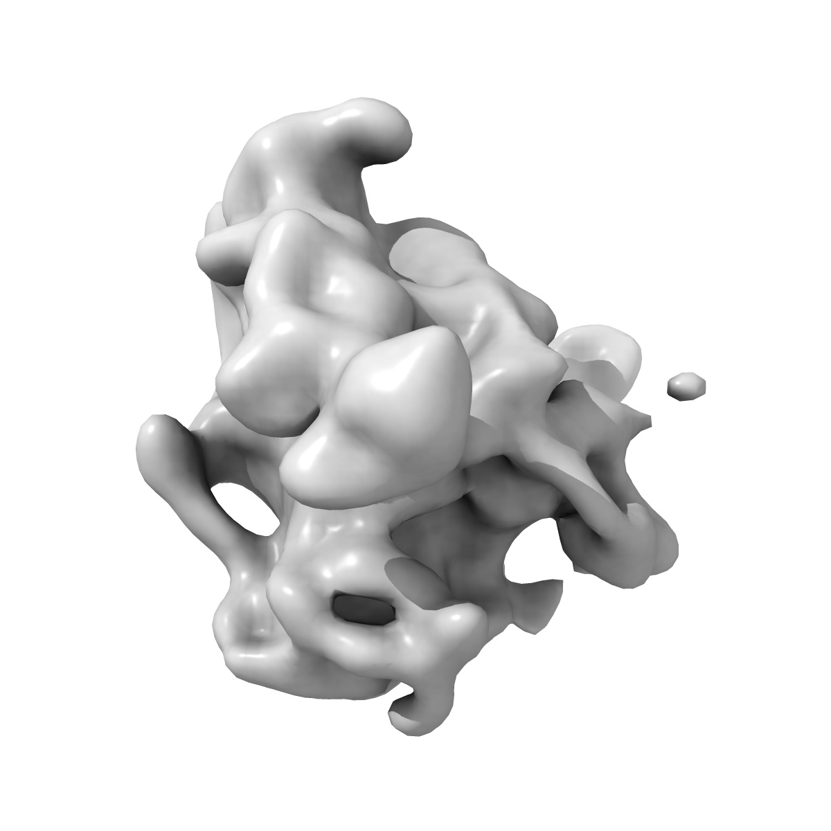
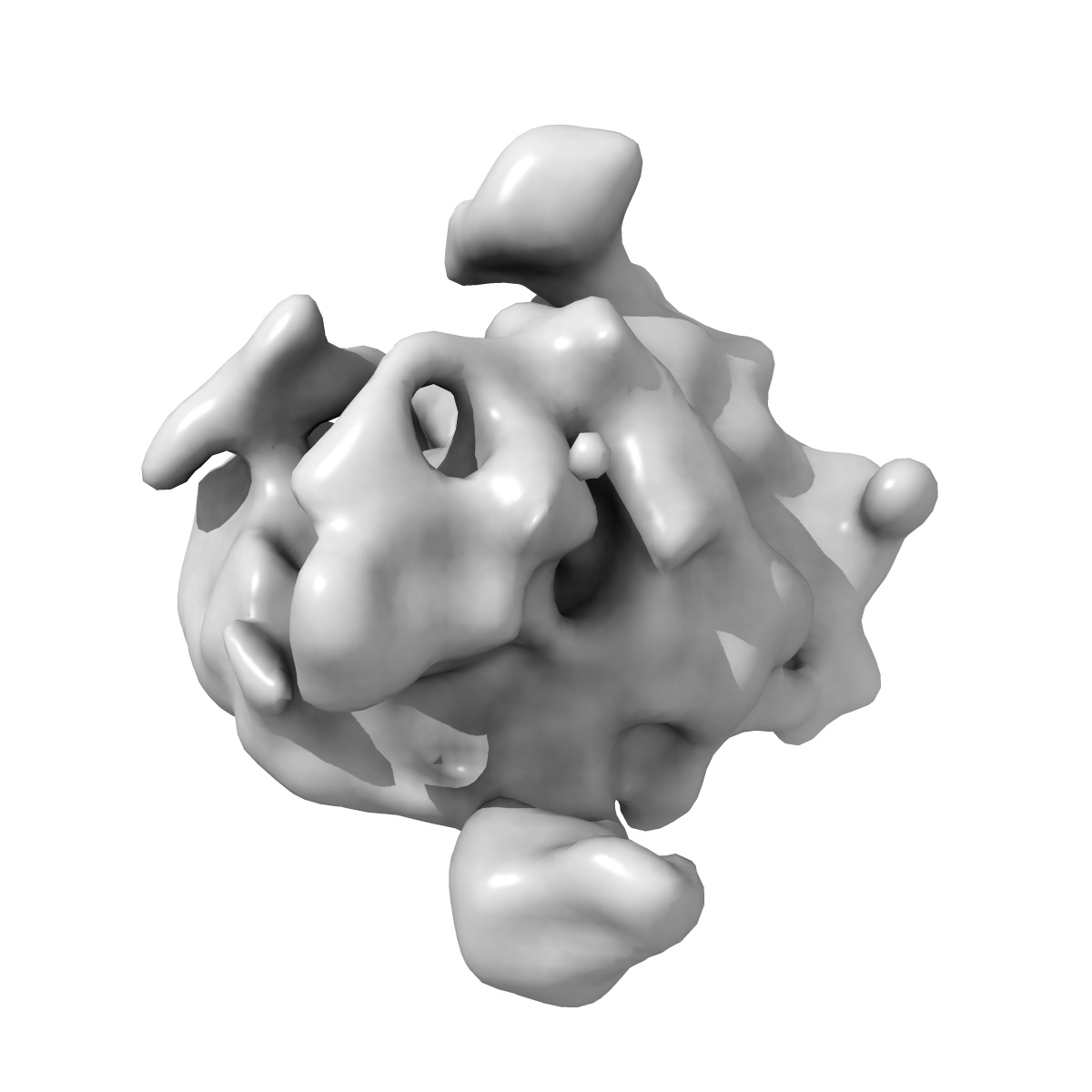
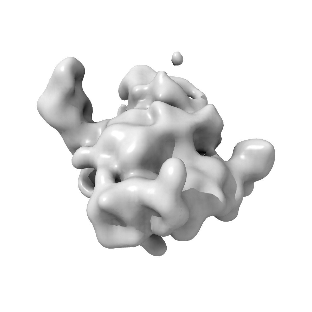
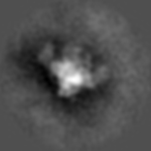
EMD-11045
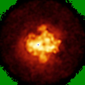
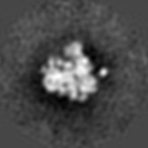
In situ cryo-electron tomography reveals layered organization of pre-ribosome maturation in nucleoli. Small Subunit Processome, Class 3
EMD-11045
Subtomogram averaging34.7 Å
 Deposition: 16/05/2020
Map released: 26/05/2021
Last modified: 13/10/2021
Sample Organism:
Chlamydomonas reinhardtii
Sample: nucleolar small subunit processome, class 1
Deposition Authors: Erdmann PS, Klumpe S, Hou Z, Beck F, Wilfling F, Plitzko JM, Baumeister W
Sample: nucleolar small subunit processome, class 1
Deposition Authors: Erdmann PS, Klumpe S, Hou Z, Beck F, Wilfling F, Plitzko JM, Baumeister W
In situ cryo-electron tomography reveals gradient organization of ribosome biogenesis in intact nucleoli.
Erdmann PS  ,
Hou Z ,
Klumpe S ,
Khavnekar S ,
Beck F,
Wilfling F ,
Plitzko JM ,
Baumeister W
,
Hou Z ,
Klumpe S ,
Khavnekar S ,
Beck F,
Wilfling F ,
Plitzko JM ,
Baumeister W
(2021) Nat Commun , 12 , 5364 - 5364
 ,
Hou Z ,
Klumpe S ,
Khavnekar S ,
Beck F,
Wilfling F ,
Plitzko JM ,
Baumeister W
,
Hou Z ,
Klumpe S ,
Khavnekar S ,
Beck F,
Wilfling F ,
Plitzko JM ,
Baumeister W
(2021) Nat Commun , 12 , 5364 - 5364
Abstract:
Ribosomes comprise a large (LSU) and a small subunit (SSU) which are synthesized independently in the nucleolus before being exported into the cytoplasm, where they assemble into functional ribosomes. Individual maturation steps have been analyzed in detail using biochemical methods, light microscopy and conventional electron microscopy (EM). In recent years, single particle analysis (SPA) has yielded molecular resolution structures of several pre-ribosomal intermediates. It falls short, however, of revealing the spatiotemporal sequence of ribosome biogenesis in the cellular context. Here, we present our study on native nucleoli in Chlamydomonas reinhardtii, in which we follow the formation of LSU and SSU precursors by in situ cryo-electron tomography (cryo-ET) and subtomogram averaging (STA). By combining both positional and molecular data, we reveal gradients of ribosome maturation within the granular component (GC), offering a new perspective on how the liquid-liquid-phase separation of the nucleolus supports ribosome biogenesis.
Ribosomes comprise a large (LSU) and a small subunit (SSU) which are synthesized independently in the nucleolus before being exported into the cytoplasm, where they assemble into functional ribosomes. Individual maturation steps have been analyzed in detail using biochemical methods, light microscopy and conventional electron microscopy (EM). In recent years, single particle analysis (SPA) has yielded molecular resolution structures of several pre-ribosomal intermediates. It falls short, however, of revealing the spatiotemporal sequence of ribosome biogenesis in the cellular context. Here, we present our study on native nucleoli in Chlamydomonas reinhardtii, in which we follow the formation of LSU and SSU precursors by in situ cryo-electron tomography (cryo-ET) and subtomogram averaging (STA). By combining both positional and molecular data, we reveal gradients of ribosome maturation within the granular component (GC), offering a new perspective on how the liquid-liquid-phase separation of the nucleolus supports ribosome biogenesis.