EMD-11383
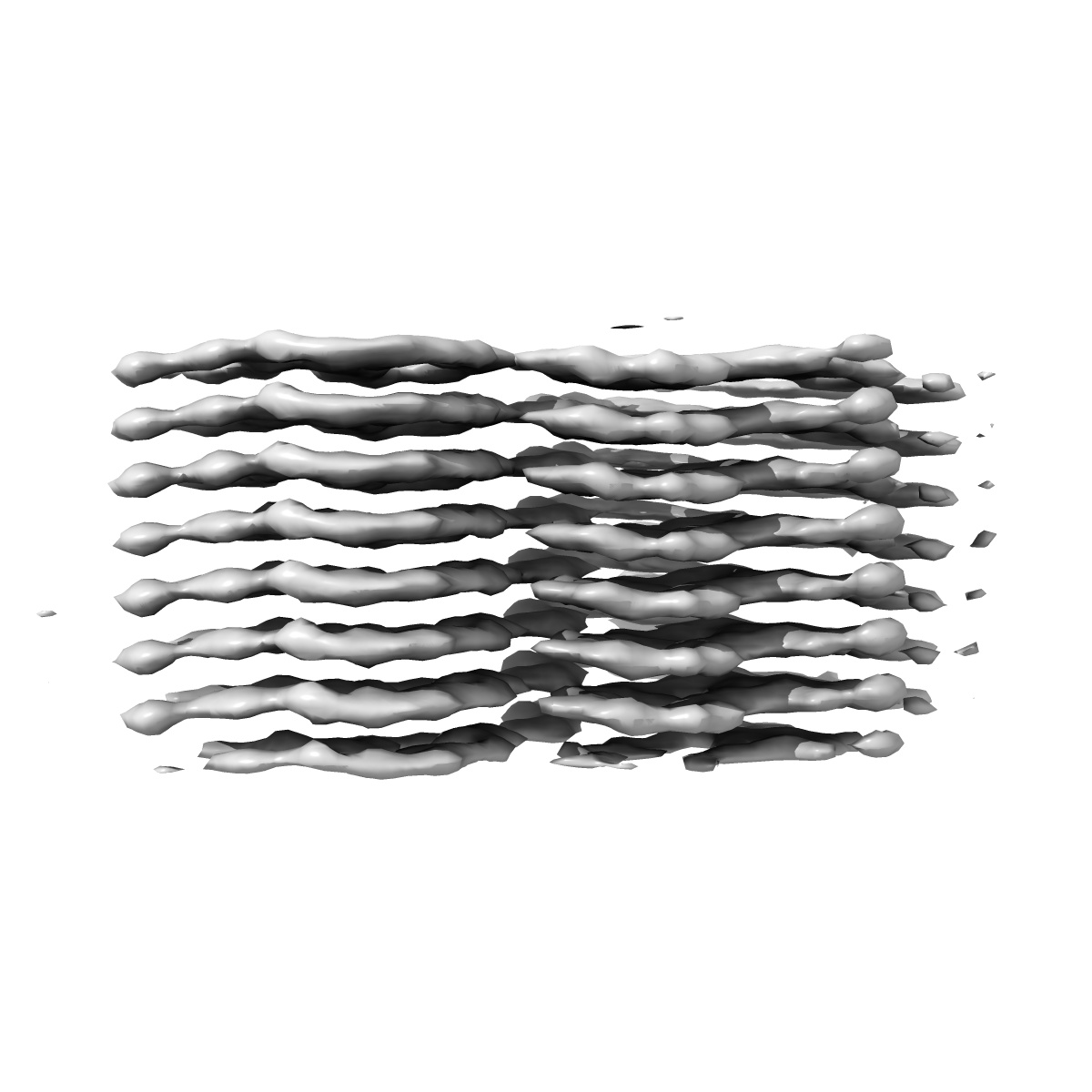
three-protofilament amyloid structure of S20G variant of human amylin (IAPP - Islet Amyloid Polypeptide)
EMD-11383
Helical reconstruction4.0 Å
 Deposition: 14/07/2020
Map released: 30/09/2020
Last modified: 18/11/2020
Sample Organism:
Homo sapiens
Sample: three-protofilament amyloid fibril of S20G variant of amylin
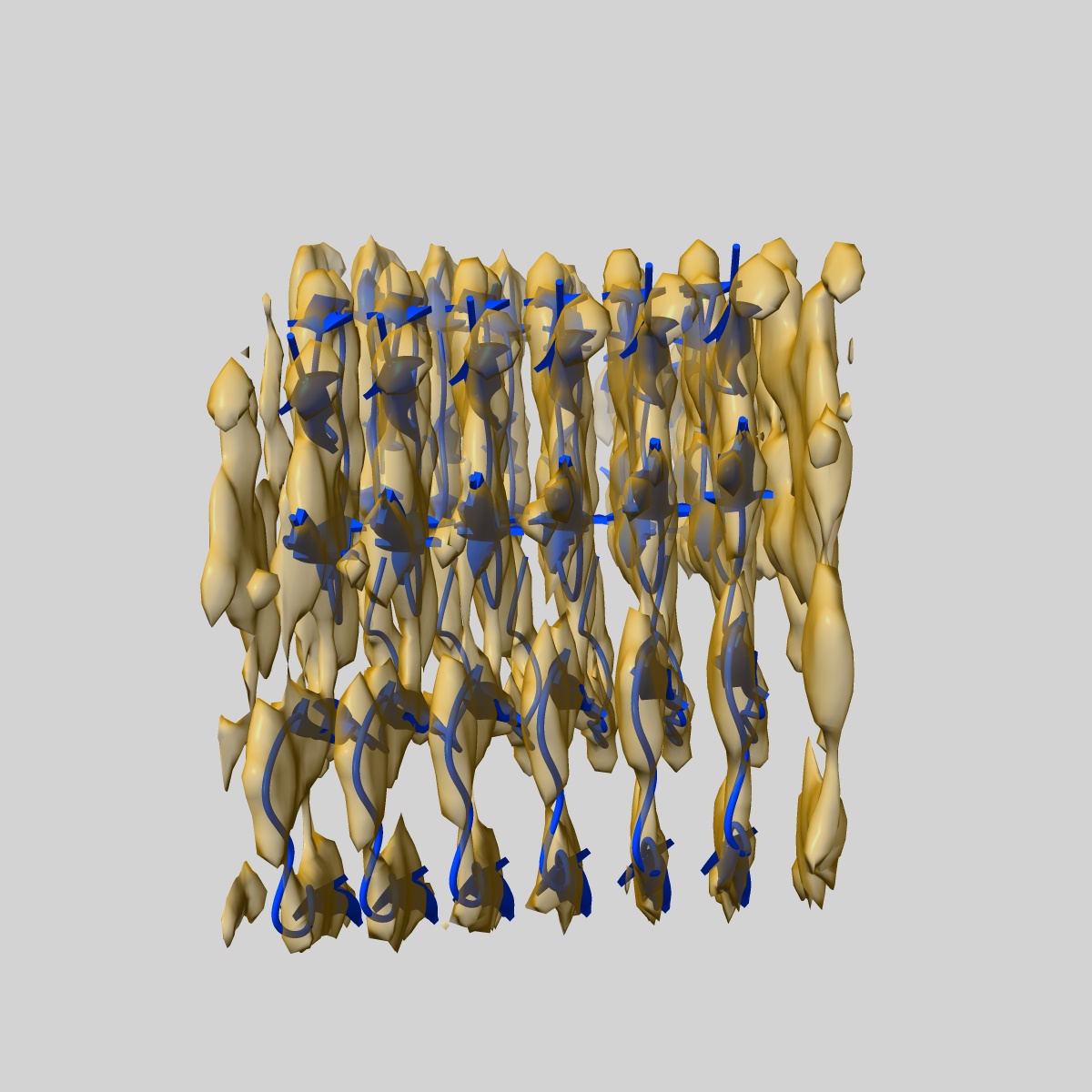
Fitted models: 6zrr (Avg. Q-score: 0.419)
Deposition Authors: Gallardo RU, Iadanza MG, Ranson NA, Radford SE
Sample: three-protofilament amyloid fibril of S20G variant of amylin
Fitted models: 6zrr (Avg. Q-score: 0.419)
Deposition Authors: Gallardo RU, Iadanza MG, Ranson NA, Radford SE
Fibril structures of diabetes-related amylin variants reveal a basis for surface-templated assembly.
Gallardo R  ,
Iadanza MG,
Xu Y ,
Heath GR ,
Foster R ,
Radford SE ,
Ranson NA
,
Iadanza MG,
Xu Y ,
Heath GR ,
Foster R ,
Radford SE ,
Ranson NA
(2020) Nat. Struct. Mol. Biol. , 27 , 1048 - 1056
 ,
Iadanza MG,
Xu Y ,
Heath GR ,
Foster R ,
Radford SE ,
Ranson NA
,
Iadanza MG,
Xu Y ,
Heath GR ,
Foster R ,
Radford SE ,
Ranson NA
(2020) Nat. Struct. Mol. Biol. , 27 , 1048 - 1056
Abstract:
Aggregation of the peptide hormone amylin into amyloid deposits is a pathological hallmark of type-2 diabetes (T2D). While no causal link between T2D and amyloid has been established, the S20G mutation in amylin is associated with early-onset T2D. Here we report cryo-EM structures of amyloid fibrils of wild-type human amylin and its S20G variant. The wild-type fibril structure, solved to 3.6-Å resolution, contains two protofilaments, each built from S-shaped subunits. S20G fibrils, by contrast, contain two major polymorphs. Their structures, solved at 3.9-Å and 4.0-Å resolution, respectively, share a common two-protofilament core that is distinct from the wild-type structure. Remarkably, one polymorph contains a third subunit with another, distinct, cross-β conformation. The presence of two different backbone conformations within the same fibril may explain the increased aggregation propensity of S20G, and illustrates a potential structural basis for surface-templated fibril assembly.
Aggregation of the peptide hormone amylin into amyloid deposits is a pathological hallmark of type-2 diabetes (T2D). While no causal link between T2D and amyloid has been established, the S20G mutation in amylin is associated with early-onset T2D. Here we report cryo-EM structures of amyloid fibrils of wild-type human amylin and its S20G variant. The wild-type fibril structure, solved to 3.6-Å resolution, contains two protofilaments, each built from S-shaped subunits. S20G fibrils, by contrast, contain two major polymorphs. Their structures, solved at 3.9-Å and 4.0-Å resolution, respectively, share a common two-protofilament core that is distinct from the wild-type structure. Remarkably, one polymorph contains a third subunit with another, distinct, cross-β conformation. The presence of two different backbone conformations within the same fibril may explain the increased aggregation propensity of S20G, and illustrates a potential structural basis for surface-templated fibril assembly.