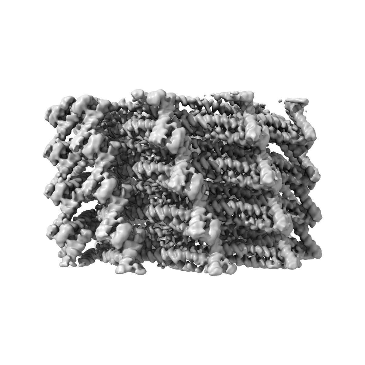
EMD-11698
Helical structure of PspA
EMD-11698
Helical reconstruction3.6 Å
 Deposition: 07/09/2020
Map released: 04/08/2021
Last modified: 10/07/2024
Sample Organism:
Synechocystis sp. (strain PCC 6803 / Kazusa)
Sample: Helical filament assembly of PspA
Fitted models: 7abk (Avg. Q-score: 0.422)
Deposition Authors: Junglas B ,
Huber ST
,
Huber ST
Sample: Helical filament assembly of PspA
Fitted models: 7abk (Avg. Q-score: 0.422)
Deposition Authors: Junglas B
 ,
Huber ST
,
Huber ST
PspA adopts an ESCRT-III-like fold and remodels bacterial membranes.
Junglas B ,
Huber ST ,
Heidler T ,
Schlosser L,
Mann D ,
Hennig R,
Clarke M ,
Hellmann N,
Schneider D,
Sachse C
(2021) Cell , 184 , 3674 - 3688.e18
,
Huber ST ,
Heidler T ,
Schlosser L,
Mann D ,
Hennig R,
Clarke M ,
Hellmann N,
Schneider D,
Sachse C
(2021) Cell , 184 , 3674 - 3688.e18
Abstract:
PspA is the main effector of the phage shock protein (Psp) system and preserves the bacterial inner membrane integrity and function. Here, we present the 3.6 Å resolution cryoelectron microscopy (cryo-EM) structure of PspA assembled in helical rods. PspA monomers adopt a canonical ESCRT-III fold in an extended open conformation. PspA rods are capable of enclosing lipids and generating positive membrane curvature. Using cryo-EM, we visualized how PspA remodels membrane vesicles into μm-sized structures and how it mediates the formation of internalized vesicular structures. Hotspots of these activities are zones derived from PspA assemblies, serving as lipid transfer platforms and linking previously separated lipid structures. These membrane fusion and fission activities are in line with the described functional properties of bacterial PspA/IM30/LiaH proteins. Our structural and functional analyses reveal that bacterial PspA belongs to the evolutionary ancestry of ESCRT-III proteins involved in membrane remodeling.
PspA is the main effector of the phage shock protein (Psp) system and preserves the bacterial inner membrane integrity and function. Here, we present the 3.6 Å resolution cryoelectron microscopy (cryo-EM) structure of PspA assembled in helical rods. PspA monomers adopt a canonical ESCRT-III fold in an extended open conformation. PspA rods are capable of enclosing lipids and generating positive membrane curvature. Using cryo-EM, we visualized how PspA remodels membrane vesicles into μm-sized structures and how it mediates the formation of internalized vesicular structures. Hotspots of these activities are zones derived from PspA assemblies, serving as lipid transfer platforms and linking previously separated lipid structures. These membrane fusion and fission activities are in line with the described functional properties of bacterial PspA/IM30/LiaH proteins. Our structural and functional analyses reveal that bacterial PspA belongs to the evolutionary ancestry of ESCRT-III proteins involved in membrane remodeling.