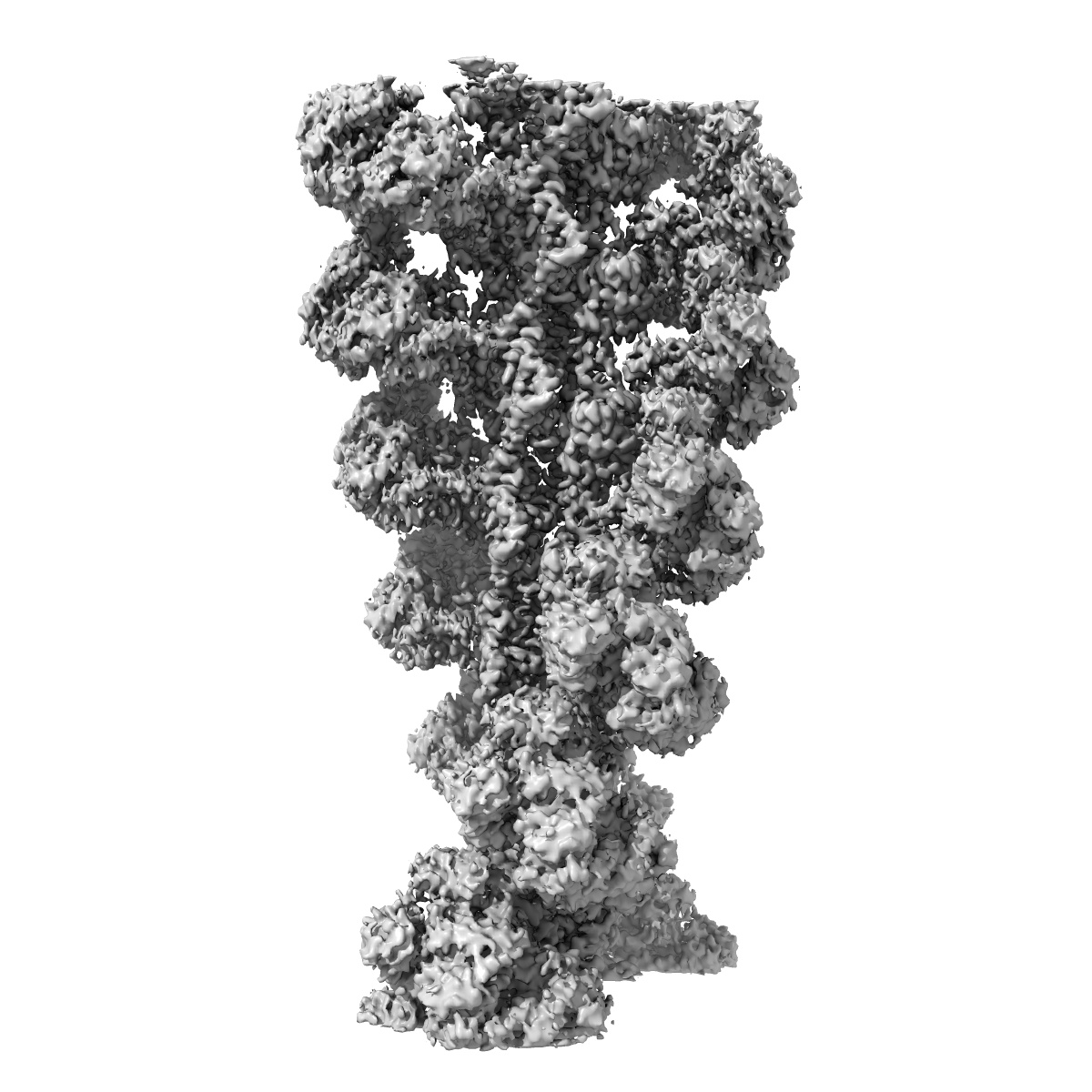
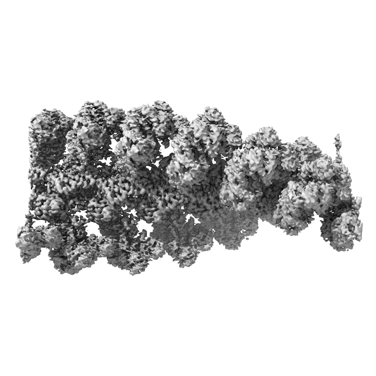
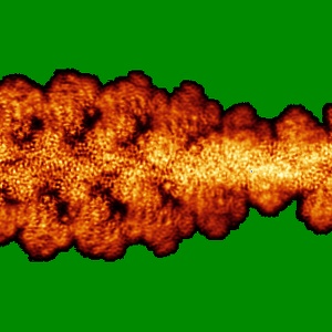
EMD-11818
Cryo-EM structure of the divergent actomyosin complex from Plasmodium falciparum Myosin A in the Rigor state
EMD-11818
Helical reconstruction3.77 Å
 Deposition: 06/10/2020
Map released: 28/04/2021
Last modified: 23/10/2024
Sample Organism:
Plasmodium falciparum (isolate 3D7)
Sample: actomyosin complex from Plasmodium falciparum
Fitted models: 7aln (Avg. Q-score: 0.402)
Deposition Authors: Robert-Paganin J, Xu X-P
Sample: actomyosin complex from Plasmodium falciparum
Fitted models: 7aln (Avg. Q-score: 0.402)
Deposition Authors: Robert-Paganin J, Xu X-P
The actomyosin interface contains an evolutionary conserved core and an ancillary interface involved in specificity.
Robert-Paganin J,
Xu XP,
Swift MF,
Auguin D  ,
Robblee JP ,
Lu H,
Fagnant PM,
Krementsova EB,
Trybus KM,
Houdusse A ,
Volkmann N ,
Hanein D
,
Robblee JP ,
Lu H,
Fagnant PM,
Krementsova EB,
Trybus KM,
Houdusse A ,
Volkmann N ,
Hanein D
(2021) Nat Commun , 12 , 1892 - 1892
 ,
Robblee JP ,
Lu H,
Fagnant PM,
Krementsova EB,
Trybus KM,
Houdusse A ,
Volkmann N ,
Hanein D
,
Robblee JP ,
Lu H,
Fagnant PM,
Krementsova EB,
Trybus KM,
Houdusse A ,
Volkmann N ,
Hanein D
(2021) Nat Commun , 12 , 1892 - 1892
Abstract:
Plasmodium falciparum, the causative agent of malaria, moves by an atypical process called gliding motility. Actomyosin interactions are central to gliding motility. However, the details of these interactions remained elusive until now. Here, we report an atomic structure of the divergent Plasmodium falciparum actomyosin system determined by electron cryomicroscopy at the end of the powerstroke (Rigor state). The structure provides insights into the detailed interactions that are required for the parasite to produce the force and motion required for infectivity. Remarkably, the footprint of the myosin motor on filamentous actin is conserved with respect to higher eukaryotes, despite important variability in the Plasmodium falciparum myosin and actin elements that make up the interface. Comparison with other actomyosin complexes reveals a conserved core interface common to all actomyosin complexes, with an ancillary interface involved in defining the spatial positioning of the motor on actin filaments.
Plasmodium falciparum, the causative agent of malaria, moves by an atypical process called gliding motility. Actomyosin interactions are central to gliding motility. However, the details of these interactions remained elusive until now. Here, we report an atomic structure of the divergent Plasmodium falciparum actomyosin system determined by electron cryomicroscopy at the end of the powerstroke (Rigor state). The structure provides insights into the detailed interactions that are required for the parasite to produce the force and motion required for infectivity. Remarkably, the footprint of the myosin motor on filamentous actin is conserved with respect to higher eukaryotes, despite important variability in the Plasmodium falciparum myosin and actin elements that make up the interface. Comparison with other actomyosin complexes reveals a conserved core interface common to all actomyosin complexes, with an ancillary interface involved in defining the spatial positioning of the motor on actin filaments.