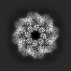
EMD-11834
hSARM1 NAD+ complex
EMD-11834
Single-particle2.68 Å
 Deposition: 13/10/2020
Map released: 11/11/2020
Last modified: 01/05/2024
Sample Organism:
Homo sapiens
Sample: hSARM1
Fitted models: 7anw (Avg. Q-score: 0.472)
Deposition Authors: Sporny M, Guez-Haddad J
Sample: hSARM1
Fitted models: 7anw (Avg. Q-score: 0.472)
Deposition Authors: Sporny M, Guez-Haddad J
Structural basis for SARM1 inhibition and activation under energetic stress.
Sporny M,
Guez-Haddad J,
Khazma T,
Yaron A  ,
Dessau M ,
Shkolnisky Y,
Mim C ,
Isupov MN ,
Zalk R,
Hons M ,
Opatowsky Y
,
Dessau M ,
Shkolnisky Y,
Mim C ,
Isupov MN ,
Zalk R,
Hons M ,
Opatowsky Y
(2020) eLife , 9
 ,
Dessau M ,
Shkolnisky Y,
Mim C ,
Isupov MN ,
Zalk R,
Hons M ,
Opatowsky Y
,
Dessau M ,
Shkolnisky Y,
Mim C ,
Isupov MN ,
Zalk R,
Hons M ,
Opatowsky Y
(2020) eLife , 9
Abstract:
SARM1, an executor of axonal degeneration, displays NADase activity that depletes the key cellular metabolite, NAD+, in response to nerve injury. The basis of SARM1 inhibition and its activation under stress conditions are still unknown. Here, we present cryo-EM maps of SARM1 at 2.9 and 2.7 Å resolutions. These indicate that SARM1 homo-octamer avoids premature activation by assuming a packed conformation, with ordered inner and peripheral rings, that prevents dimerization and activation of the catalytic domains. This inactive conformation is stabilized by binding of SARM1's own substrate NAD+ in an allosteric location, away from the catalytic sites. This model was validated by mutagenesis of the allosteric site, which led to constitutively active SARM1. We propose that the reduction of cellular NAD+ concentration contributes to the disassembly of SARM1's peripheral ring, which allows formation of active NADase domain dimers, thereby further depleting NAD+ to cause an energetic catastrophe and cell death.
SARM1, an executor of axonal degeneration, displays NADase activity that depletes the key cellular metabolite, NAD+, in response to nerve injury. The basis of SARM1 inhibition and its activation under stress conditions are still unknown. Here, we present cryo-EM maps of SARM1 at 2.9 and 2.7 Å resolutions. These indicate that SARM1 homo-octamer avoids premature activation by assuming a packed conformation, with ordered inner and peripheral rings, that prevents dimerization and activation of the catalytic domains. This inactive conformation is stabilized by binding of SARM1's own substrate NAD+ in an allosteric location, away from the catalytic sites. This model was validated by mutagenesis of the allosteric site, which led to constitutively active SARM1. We propose that the reduction of cellular NAD+ concentration contributes to the disassembly of SARM1's peripheral ring, which allows formation of active NADase domain dimers, thereby further depleting NAD+ to cause an energetic catastrophe and cell death.