EMD-11938
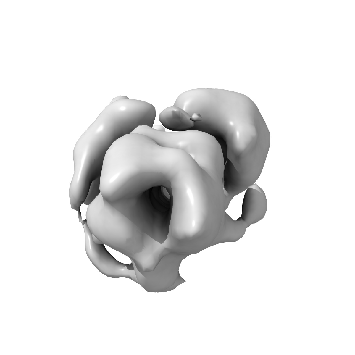
Ternary complex of full-length Caspase-8 and FADD
EMD-11938
Single-particle22.41 Å
 Deposition: 16/11/2020
Map released: 17/02/2021
Last modified: 17/02/2021
Sample Organism:
Homo sapiens
Sample: Ternary complex of full-length Caspase-8 with FADD
Deposition Authors: Fox JL, Ragan TJ, Dinsdale D, Fairall L, Schwabe JWR, Morone N, Cain K, MacFarlane M
Sample: Ternary complex of full-length Caspase-8 with FADD
Deposition Authors: Fox JL, Ragan TJ, Dinsdale D, Fairall L, Schwabe JWR, Morone N, Cain K, MacFarlane M
Cryo-EM structural analysis of FADD:Caspase-8 complexes defines the catalytic dimer architecture for co-ordinated control of cell fate.
Fox JL  ,
Hughes MA,
Meng X,
Sarnowska NA,
Powley IR,
Jukes-Jones R,
Dinsdale D,
Ragan TJ ,
Fairall L ,
Schwabe JWR ,
Morone N ,
Cain K,
MacFarlane M
,
Hughes MA,
Meng X,
Sarnowska NA,
Powley IR,
Jukes-Jones R,
Dinsdale D,
Ragan TJ ,
Fairall L ,
Schwabe JWR ,
Morone N ,
Cain K,
MacFarlane M
(2021) Nat Commun , 12 , 819 - 819
 ,
Hughes MA,
Meng X,
Sarnowska NA,
Powley IR,
Jukes-Jones R,
Dinsdale D,
Ragan TJ ,
Fairall L ,
Schwabe JWR ,
Morone N ,
Cain K,
MacFarlane M
,
Hughes MA,
Meng X,
Sarnowska NA,
Powley IR,
Jukes-Jones R,
Dinsdale D,
Ragan TJ ,
Fairall L ,
Schwabe JWR ,
Morone N ,
Cain K,
MacFarlane M
(2021) Nat Commun , 12 , 819 - 819
Abstract:
Regulated cell death is essential in development and cellular homeostasis. Multi-protein platforms, including the Death-Inducing Signaling Complex (DISC), co-ordinate cell fate via a core FADD:Caspase-8 complex and its regulatory partners, such as the cell death inhibitor c-FLIP. Here, using electron microscopy, we visualize full-length procaspase-8 in complex with FADD. Our structural analysis now reveals how the FADD-nucleated tandem death effector domain (tDED) helical filament is required to orientate the procaspase-8 catalytic domains, enabling their activation via anti-parallel dimerization. Strikingly, recruitment of c-FLIPS into this complex inhibits Caspase-8 activity by altering tDED triple helix architecture, resulting in steric hindrance of the canonical tDED Type I binding site. This prevents both Caspase-8 catalytic domain assembly and tDED helical filament elongation. Our findings reveal how the plasticity, composition and architecture of the core FADD:Caspase-8 complex critically defines life/death decisions not only via the DISC, but across multiple key signaling platforms including TNF complex II, the ripoptosome, and RIPK1/RIPK3 necrosome.
Regulated cell death is essential in development and cellular homeostasis. Multi-protein platforms, including the Death-Inducing Signaling Complex (DISC), co-ordinate cell fate via a core FADD:Caspase-8 complex and its regulatory partners, such as the cell death inhibitor c-FLIP. Here, using electron microscopy, we visualize full-length procaspase-8 in complex with FADD. Our structural analysis now reveals how the FADD-nucleated tandem death effector domain (tDED) helical filament is required to orientate the procaspase-8 catalytic domains, enabling their activation via anti-parallel dimerization. Strikingly, recruitment of c-FLIPS into this complex inhibits Caspase-8 activity by altering tDED triple helix architecture, resulting in steric hindrance of the canonical tDED Type I binding site. This prevents both Caspase-8 catalytic domain assembly and tDED helical filament elongation. Our findings reveal how the plasticity, composition and architecture of the core FADD:Caspase-8 complex critically defines life/death decisions not only via the DISC, but across multiple key signaling platforms including TNF complex II, the ripoptosome, and RIPK1/RIPK3 necrosome.