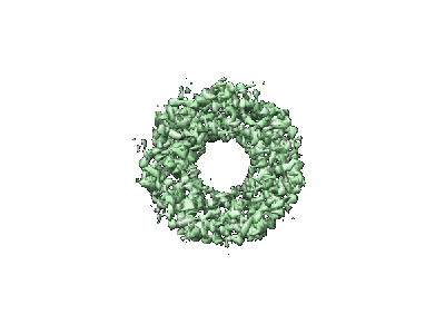
EMD-1200
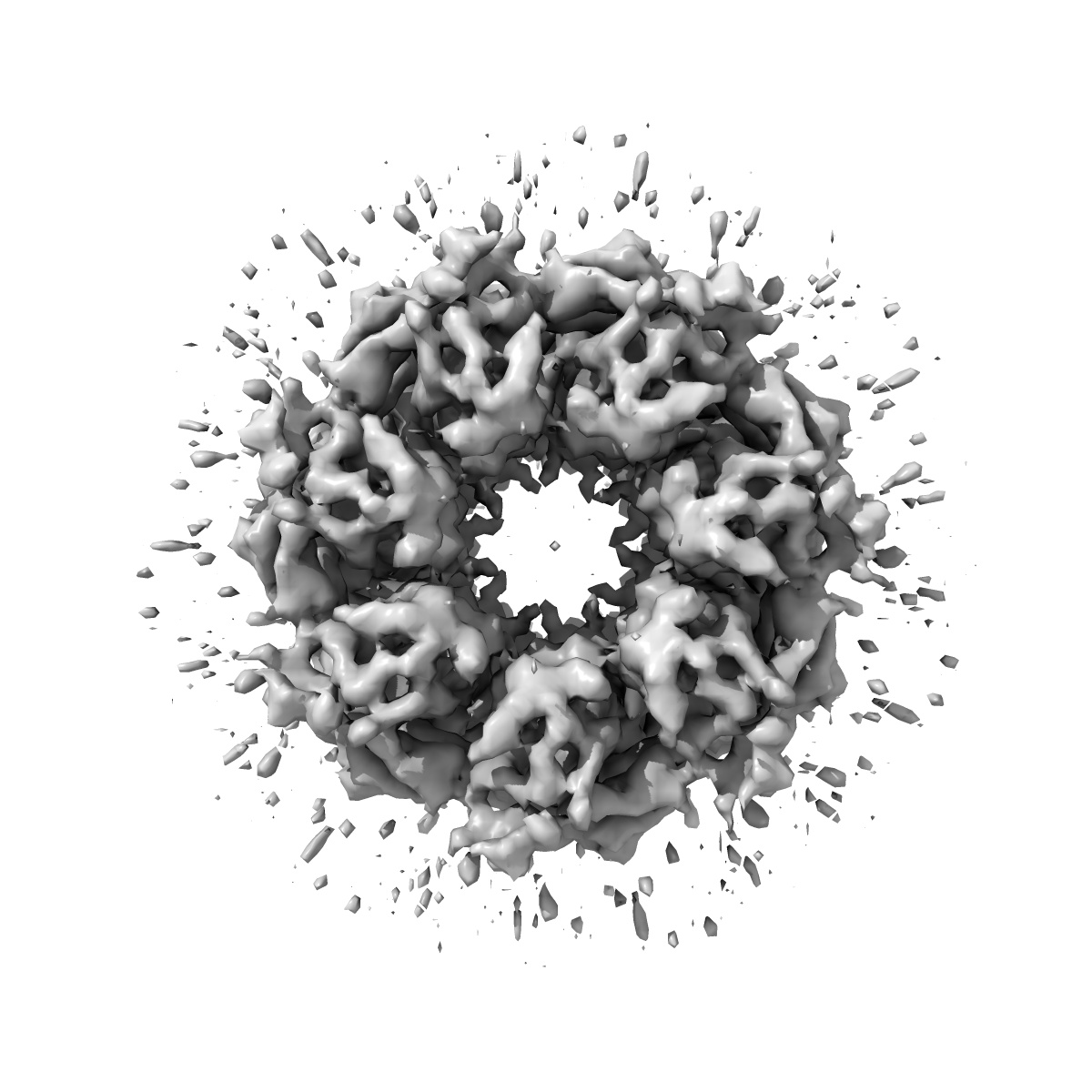
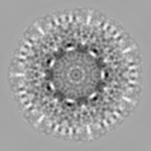
Automated cryoEM data acquisition and analysis of 284742 particles of GroEL.
EMD-1200
Single-particle7.8 Å
 Deposition: 28/02/2006
Map released: 01/07/2006
Last modified: 26/05/2011
Sample Organism:
Escherichia coli
Sample: E.coli GroEL
Raw data: EMPIAR-10018
Deposition Authors: Stagg SM ,
Pulokas J,
Fellmann D,
Cheng A,
Quispe JD ,
Mallick SP,
Avila RM,
Carragher B ,
Potter CS
,
Pulokas J,
Fellmann D,
Cheng A,
Quispe JD ,
Mallick SP,
Avila RM,
Carragher B ,
Potter CS
Sample: E.coli GroEL
Raw data: EMPIAR-10018
Deposition Authors: Stagg SM
 ,
Pulokas J,
Fellmann D,
Cheng A,
Quispe JD ,
Mallick SP,
Avila RM,
Carragher B ,
Potter CS
,
Pulokas J,
Fellmann D,
Cheng A,
Quispe JD ,
Mallick SP,
Avila RM,
Carragher B ,
Potter CS
Automated cryoEM data acquisition and analysis of 284742 particles of GroEL.
Stagg SM ,
Lander GC ,
Pulokas J,
Fellmann D,
Cheng A,
Quispe JD ,
Mallick SP,
Avila RM,
Carragher B ,
Potter CS
(2006) J Struct Biol , 155 , 470 - 481
,
Lander GC ,
Pulokas J,
Fellmann D,
Cheng A,
Quispe JD ,
Mallick SP,
Avila RM,
Carragher B ,
Potter CS
(2006) J Struct Biol , 155 , 470 - 481
Abstract:
One of the goals in developing our automated electron microscopy data acquisition system, Leginon, was to improve both the ease of use and the throughput of the process of acquiring low dose images of macromolecular specimens embedded in vitreous ice. In this article, we demonstrate the potential of the Leginon system for high-throughput data acquisition by describing an experiment in which we acquired images of more than 280,000 particles of GroEL in a single 25 h session at the microscope. We also demonstrate the potential for an automated pipeline for molecular microscopy by showing that these particles can be subjected to completely automated procedures to reconstruct a three-dimensional (3D) density map to a resolution better than 8 A. In generating the 3D maps, we used a variety of metadata associated with the data acquisition and processing steps to sort and select the particles. These metadata provide a number of insights into factors that affect the quality of the acquired images and the resulting reconstructions. In particular, we show that the resolution of the reconstructed 3D density maps improves with decreasing ice thickness. These data provide a basis for assessing the capabilities of high-throughput macromolecular microscopy.
One of the goals in developing our automated electron microscopy data acquisition system, Leginon, was to improve both the ease of use and the throughput of the process of acquiring low dose images of macromolecular specimens embedded in vitreous ice. In this article, we demonstrate the potential of the Leginon system for high-throughput data acquisition by describing an experiment in which we acquired images of more than 280,000 particles of GroEL in a single 25 h session at the microscope. We also demonstrate the potential for an automated pipeline for molecular microscopy by showing that these particles can be subjected to completely automated procedures to reconstruct a three-dimensional (3D) density map to a resolution better than 8 A. In generating the 3D maps, we used a variety of metadata associated with the data acquisition and processing steps to sort and select the particles. These metadata provide a number of insights into factors that affect the quality of the acquired images and the resulting reconstructions. In particular, we show that the resolution of the reconstructed 3D density maps improves with decreasing ice thickness. These data provide a basis for assessing the capabilities of high-throughput macromolecular microscopy.