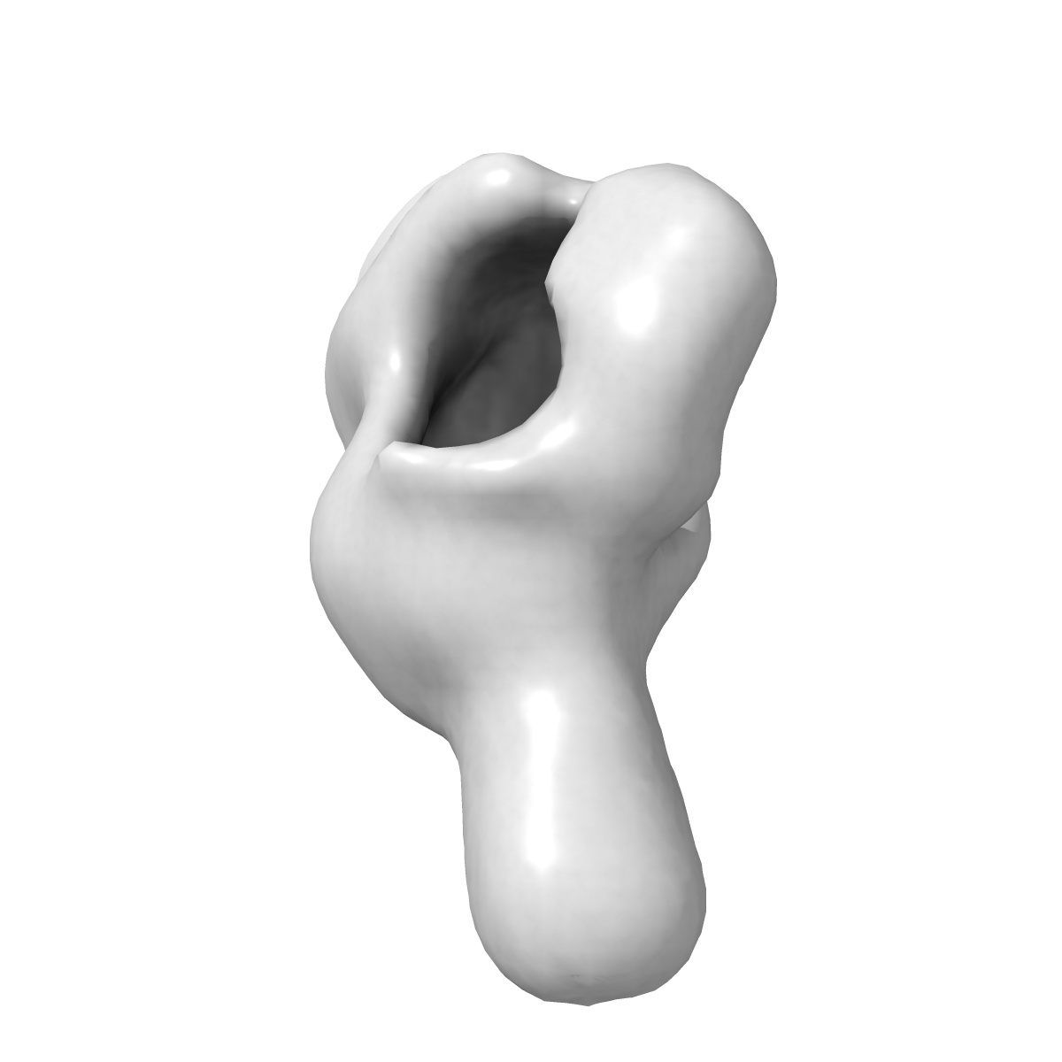
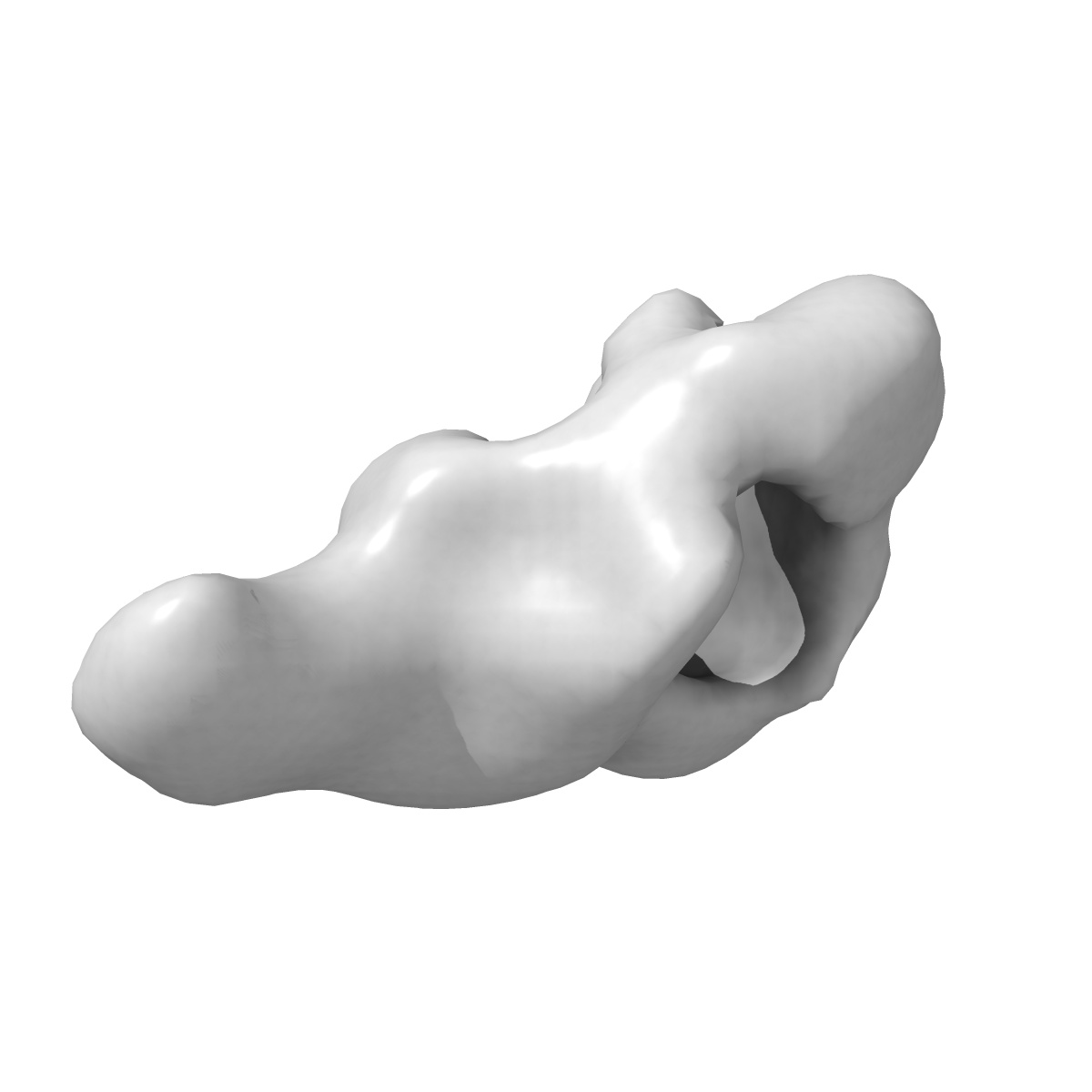
EMD-13151
Negative stain EM 3D Reconstruction of the Dam1cBim1p complex.
EMD-13151
Single-particle35.0 Å
 Deposition: 30/06/2021
Map released: 28/07/2021
Last modified: 29/09/2021
Sample Organism:
Saccharomyces cerevisiae
Sample: Dam1pBim1p Complex
Deposition Authors: Engelhard L, Bourque CB, Klink BU, Gatsogiannis C
Sample: Dam1pBim1p Complex
Deposition Authors: Engelhard L, Bourque CB, Klink BU, Gatsogiannis C
Phospho-regulated Bim1/EB1 interactions trigger Dam1c ring assembly at the budding yeast outer kinetochore.
Dudziak A  ,
Engelhard L,
Bourque C,
Klink BU,
Rombaut P,
Kornakov N ,
Janen K,
Herzog F ,
Gatsogiannis C,
Westermann S
,
Engelhard L,
Bourque C,
Klink BU,
Rombaut P,
Kornakov N ,
Janen K,
Herzog F ,
Gatsogiannis C,
Westermann S
(2021) Embo J. , 40 , e108004 - e108004
 ,
Engelhard L,
Bourque C,
Klink BU,
Rombaut P,
Kornakov N ,
Janen K,
Herzog F ,
Gatsogiannis C,
Westermann S
,
Engelhard L,
Bourque C,
Klink BU,
Rombaut P,
Kornakov N ,
Janen K,
Herzog F ,
Gatsogiannis C,
Westermann S
(2021) Embo J. , 40 , e108004 - e108004
Abstract:
Kinetochores form the link between chromosomes and microtubules of the mitotic spindle. The heterodecameric Dam1 complex (Dam1c) is a major component of the Saccharomyces cerevisiae outer kinetochore, assembling into 3 MDa-sized microtubule-embracing rings, but how ring assembly is specifically initiated in vivo remains to be understood. Here, we describe a molecular pathway that provides local control of ring assembly during the establishment of sister kinetochore bi-orientation. We show that Dam1c and the general microtubule plus end-associated protein (+TIP) Bim1/EB1 form a stable complex depending on a conserved motif in the Duo1 subunit of Dam1c. EM analyses reveal that Bim1 crosslinks protrusion domains of adjacent Dam1c heterodecamers and promotes the formation of oligomers with defined curvature. Disruption of the Dam1c-Bim1 interaction impairs kinetochore localization of Dam1c in metaphase and delays mitosis. Phosphorylation promotes Dam1c-Bim1 binding by relieving an intramolecular inhibition of the Dam1 C-terminus. In addition, Bim1 recruits Bik1/CLIP-170 to Dam1c and induces formation of full rings even in the absence of microtubules. Our data help to explain how new kinetochore end-on attachments are formed during the process of attachment error correction.
Kinetochores form the link between chromosomes and microtubules of the mitotic spindle. The heterodecameric Dam1 complex (Dam1c) is a major component of the Saccharomyces cerevisiae outer kinetochore, assembling into 3 MDa-sized microtubule-embracing rings, but how ring assembly is specifically initiated in vivo remains to be understood. Here, we describe a molecular pathway that provides local control of ring assembly during the establishment of sister kinetochore bi-orientation. We show that Dam1c and the general microtubule plus end-associated protein (+TIP) Bim1/EB1 form a stable complex depending on a conserved motif in the Duo1 subunit of Dam1c. EM analyses reveal that Bim1 crosslinks protrusion domains of adjacent Dam1c heterodecamers and promotes the formation of oligomers with defined curvature. Disruption of the Dam1c-Bim1 interaction impairs kinetochore localization of Dam1c in metaphase and delays mitosis. Phosphorylation promotes Dam1c-Bim1 binding by relieving an intramolecular inhibition of the Dam1 C-terminus. In addition, Bim1 recruits Bik1/CLIP-170 to Dam1c and induces formation of full rings even in the absence of microtubules. Our data help to explain how new kinetochore end-on attachments are formed during the process of attachment error correction.