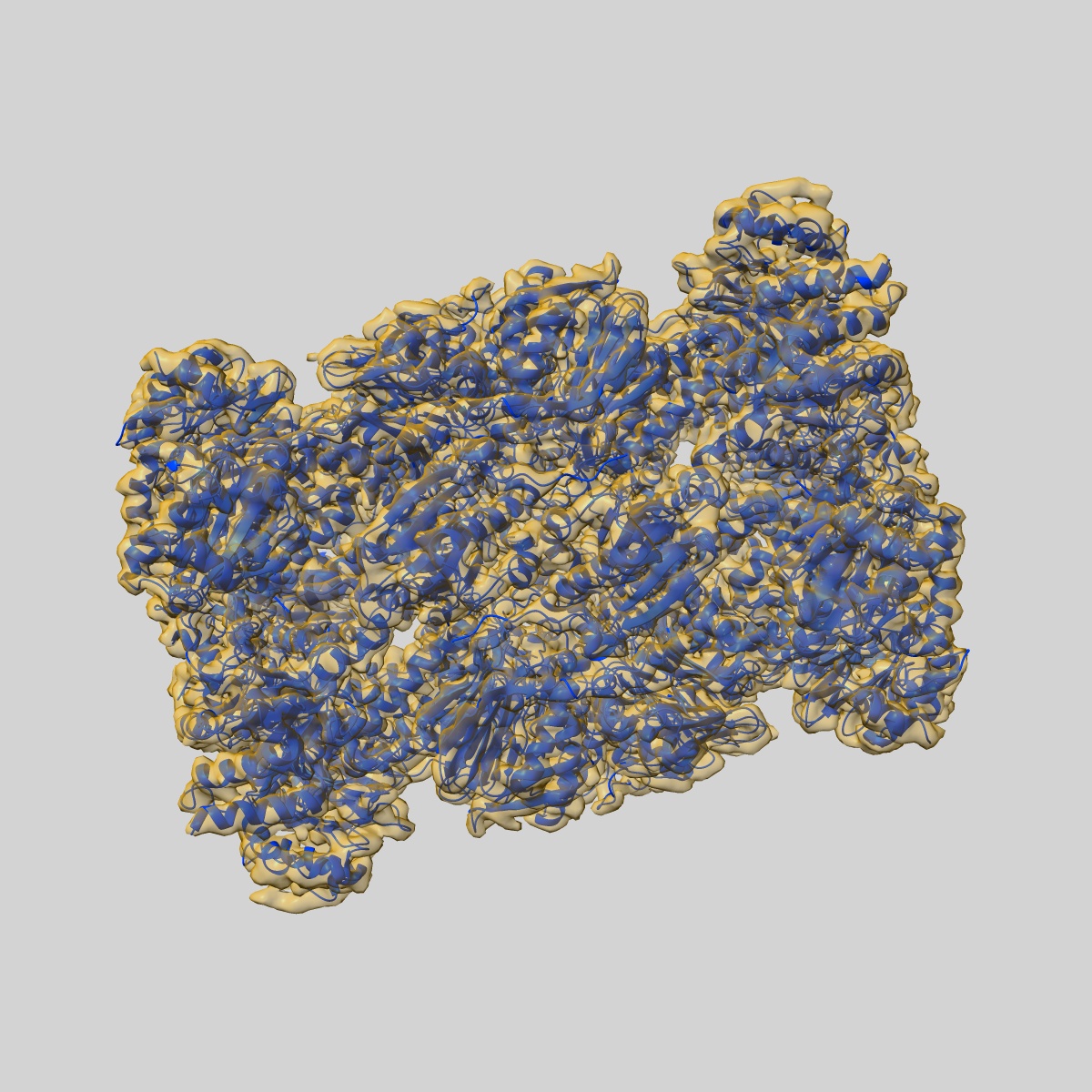
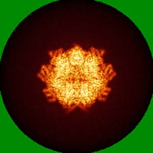
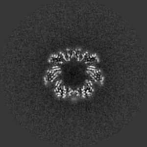
EMD-13389
human 20S proteasome (before post-processing)
EMD-13389
Single-particle3.7 Å
 Deposition: 13/08/2021
Map released: 20/10/2021
Last modified: 17/07/2024
Sample Organism:
Homo sapiens
Sample: human 20S proteasome
Fitted models: 7pg9 (Avg. Q-score: 0.444)
Deposition Authors: Xu C, Cong Y
Sample: human 20S proteasome
Fitted models: 7pg9 (Avg. Q-score: 0.444)
Deposition Authors: Xu C, Cong Y

The 20S as a stand-alone proteasome in cells can degrade the ubiquitin tag.
Sahu I ,
Mali SM,
Sulkshane P ,
Xu C,
Rozenberg A,
Morag R,
Sahoo MP,
Singh SK ,
Ding Z,
Wang Y ,
Day S,
Cong Y ,
Kleifeld O ,
Brik A ,
Glickman MH
(2021) Nat Commun , 12 , 6173 - 6173
,
Mali SM,
Sulkshane P ,
Xu C,
Rozenberg A,
Morag R,
Sahoo MP,
Singh SK ,
Ding Z,
Wang Y ,
Day S,
Cong Y ,
Kleifeld O ,
Brik A ,
Glickman MH
(2021) Nat Commun , 12 , 6173 - 6173
Abstract:
The proteasome, the primary protease for ubiquitin-dependent proteolysis in eukaryotes, is usually found as a mixture of 30S, 26S, and 20S complexes. These complexes have common catalytic sites, which makes it challenging to determine their distinctive roles in intracellular proteolysis. Here, we chemically synthesize a panel of homogenous ubiquitinated proteins, and use them to compare 20S and 26S proteasomes with respect to substrate selection and peptide-product generation. We show that 20S proteasomes can degrade the ubiquitin tag along with the conjugated substrate. Ubiquitin remnants on branched peptide products identified by LC-MS/MS, and flexibility in the 20S gate observed by cryo-EM, reflect the ability of the 20S proteasome to proteolyze an isopeptide-linked ubiquitin-conjugate. Peptidomics identifies proteasome-trapped ubiquitin-derived peptides and peptides of potential 20S substrates in Hi20S cells, hypoxic cells, and human failing-heart. Moreover, elevated levels of 20S proteasomes appear to contribute to cell survival under stress associated with damaged proteins.
The proteasome, the primary protease for ubiquitin-dependent proteolysis in eukaryotes, is usually found as a mixture of 30S, 26S, and 20S complexes. These complexes have common catalytic sites, which makes it challenging to determine their distinctive roles in intracellular proteolysis. Here, we chemically synthesize a panel of homogenous ubiquitinated proteins, and use them to compare 20S and 26S proteasomes with respect to substrate selection and peptide-product generation. We show that 20S proteasomes can degrade the ubiquitin tag along with the conjugated substrate. Ubiquitin remnants on branched peptide products identified by LC-MS/MS, and flexibility in the 20S gate observed by cryo-EM, reflect the ability of the 20S proteasome to proteolyze an isopeptide-linked ubiquitin-conjugate. Peptidomics identifies proteasome-trapped ubiquitin-derived peptides and peptides of potential 20S substrates in Hi20S cells, hypoxic cells, and human failing-heart. Moreover, elevated levels of 20S proteasomes appear to contribute to cell survival under stress associated with damaged proteins.