EMD-14221
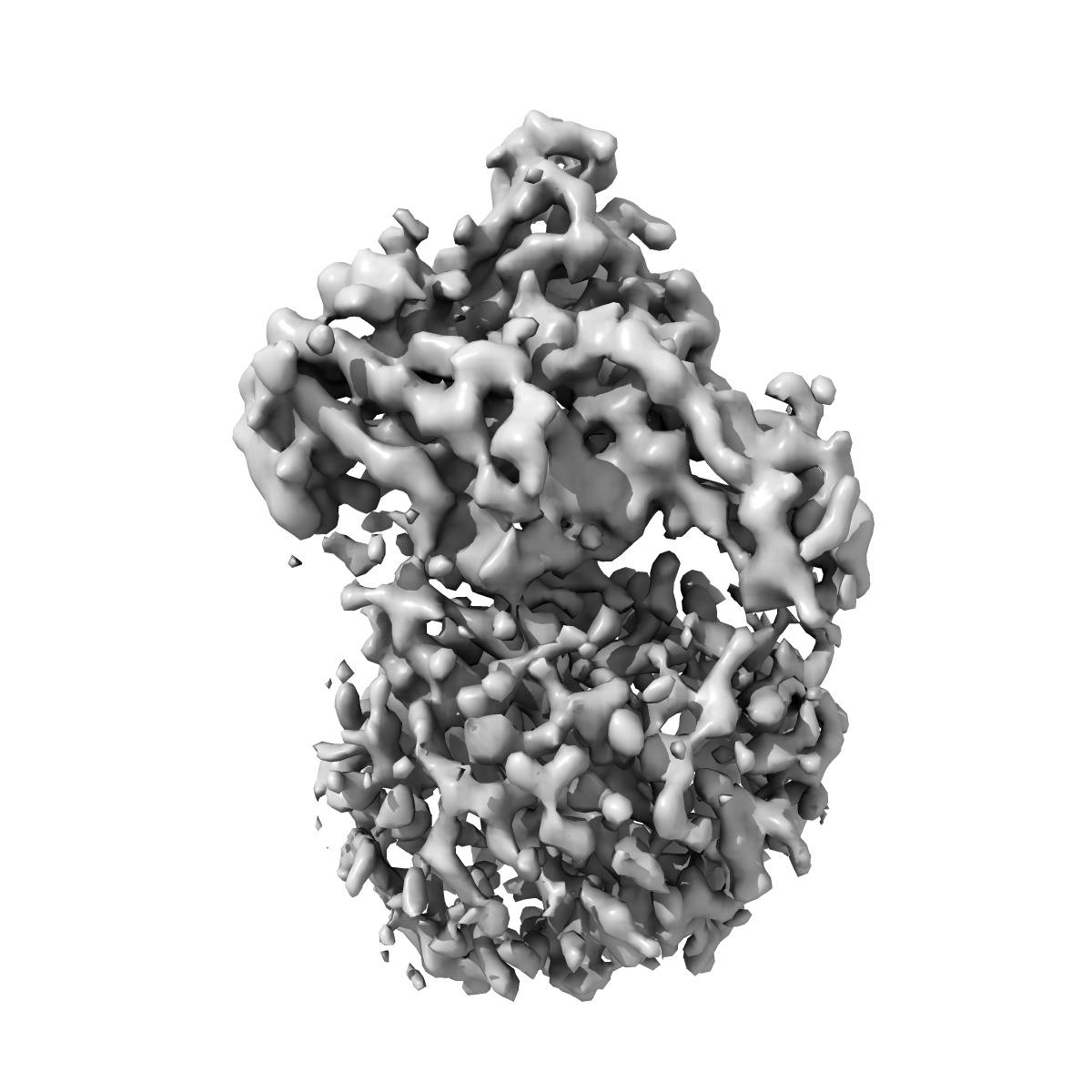
Structure of the AVP-V2R-arrestin2-ScFv30 complex
EMD-14221
Single-particle4.73 Å
 Deposition: 01/02/2022
Map released: 14/09/2022
Last modified: 23/10/2024
Sample Organism:
Homo sapiens,
synthetic construct
Sample: Ternary complex of the AVP-V2 receptor with arrestin2 and ScfV30
Fitted models: 7r0c (Avg. Q-score: 0.249)
Deposition Authors: Bous J ,
Fouillen A ,
Trapani S ,
Granier S ,
Mouillac B ,
Bron P
,
Fouillen A ,
Trapani S ,
Granier S ,
Mouillac B ,
Bron P
Sample: Ternary complex of the AVP-V2 receptor with arrestin2 and ScfV30
Fitted models: 7r0c (Avg. Q-score: 0.249)
Deposition Authors: Bous J
 ,
Fouillen A ,
Trapani S ,
Granier S ,
Mouillac B ,
Bron P
,
Fouillen A ,
Trapani S ,
Granier S ,
Mouillac B ,
Bron P
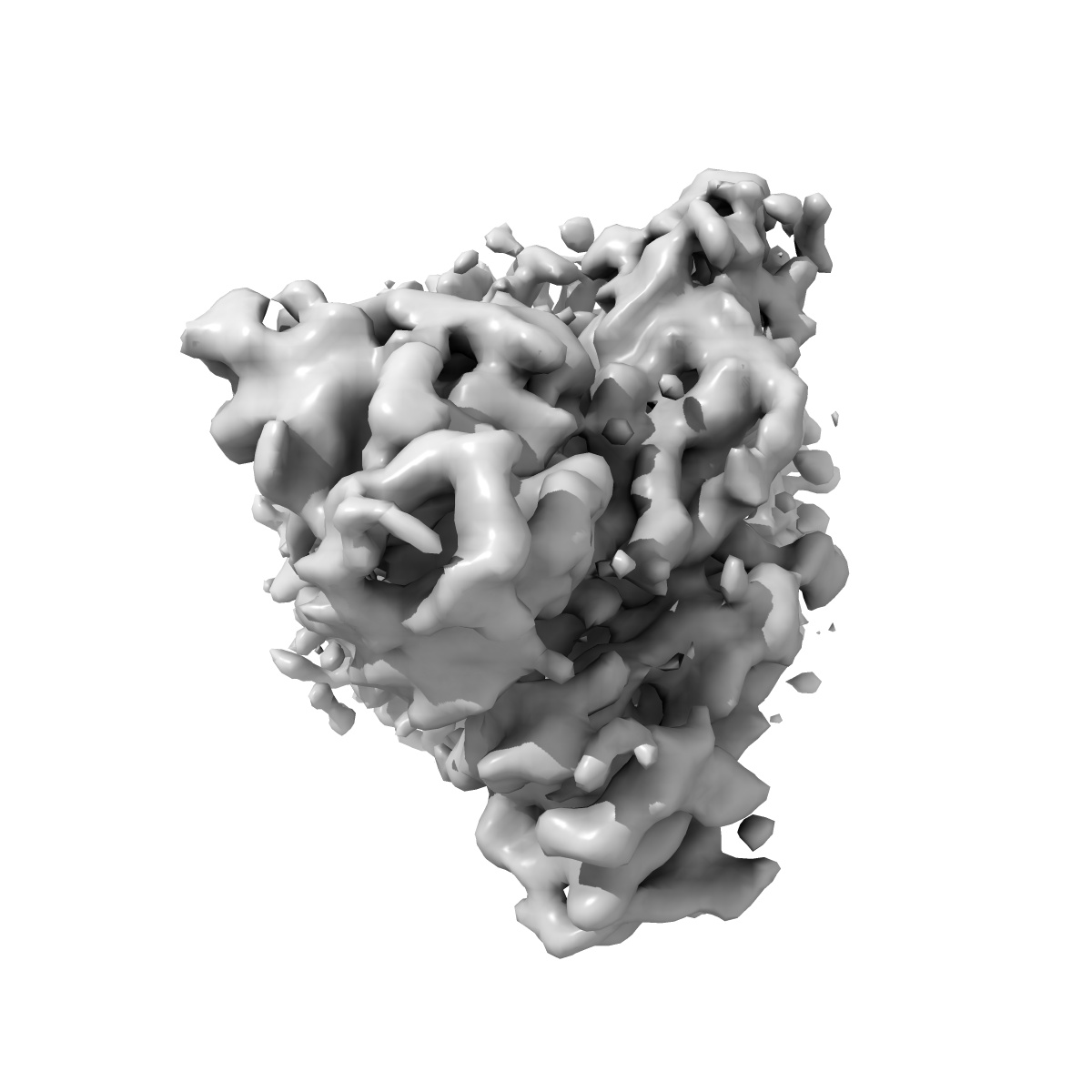
Structure of the vasopressin hormone-V2 receptor-beta-arrestin1 ternary complex.
Bous J ,
Fouillen A ,
Orcel H ,
Trapani S ,
Cong X ,
Fontanel S ,
Saint-Paul J ,
Lai-Kee-Him J,
Urbach S ,
Sibille N ,
Sounier R ,
Granier S ,
Mouillac B ,
Bron P
(2022) Sci Adv , 8 , eabo7761 - eabo7761
,
Fouillen A ,
Orcel H ,
Trapani S ,
Cong X ,
Fontanel S ,
Saint-Paul J ,
Lai-Kee-Him J,
Urbach S ,
Sibille N ,
Sounier R ,
Granier S ,
Mouillac B ,
Bron P
(2022) Sci Adv , 8 , eabo7761 - eabo7761
Abstract:
Arrestins interact with G protein-coupled receptors (GPCRs) to stop G protein activation and to initiate key signaling pathways. Recent structural studies shed light on the molecular mechanisms involved in GPCR-arrestin coupling, but whether this process is conserved among GPCRs is poorly understood. Here, we report the cryo-electron microscopy active structure of the wild-type arginine-vasopressin V2 receptor (V2R) in complex with β-arrestin1. It reveals an atypical position of β-arrestin1 compared to previously described GPCR-arrestin assemblies, associated with an original V2R/β-arrestin1 interface involving all receptor intracellular loops. Phosphorylated sites of the V2R carboxyl terminus are clearly identified and interact extensively with the β-arrestin1 N-lobe, in agreement with structural data obtained with chimeric or synthetic systems. Overall, these findings highlight a notable structural variability among GPCR-arrestin signaling complexes.
Arrestins interact with G protein-coupled receptors (GPCRs) to stop G protein activation and to initiate key signaling pathways. Recent structural studies shed light on the molecular mechanisms involved in GPCR-arrestin coupling, but whether this process is conserved among GPCRs is poorly understood. Here, we report the cryo-electron microscopy active structure of the wild-type arginine-vasopressin V2 receptor (V2R) in complex with β-arrestin1. It reveals an atypical position of β-arrestin1 compared to previously described GPCR-arrestin assemblies, associated with an original V2R/β-arrestin1 interface involving all receptor intracellular loops. Phosphorylated sites of the V2R carboxyl terminus are clearly identified and interact extensively with the β-arrestin1 N-lobe, in agreement with structural data obtained with chimeric or synthetic systems. Overall, these findings highlight a notable structural variability among GPCR-arrestin signaling complexes.