EMD-1459
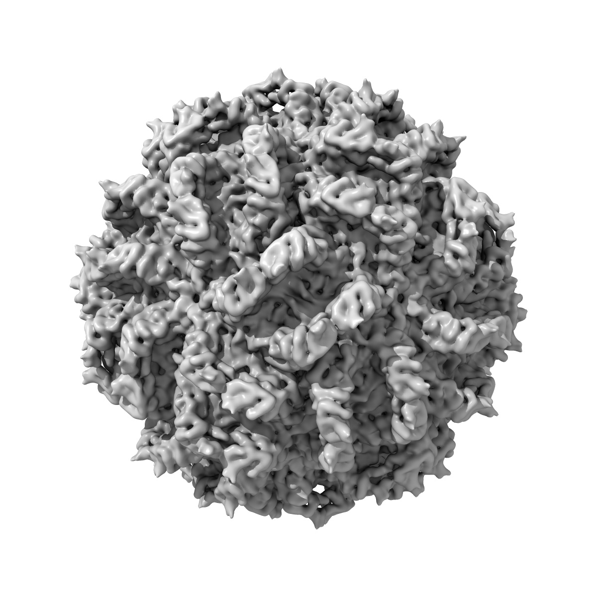
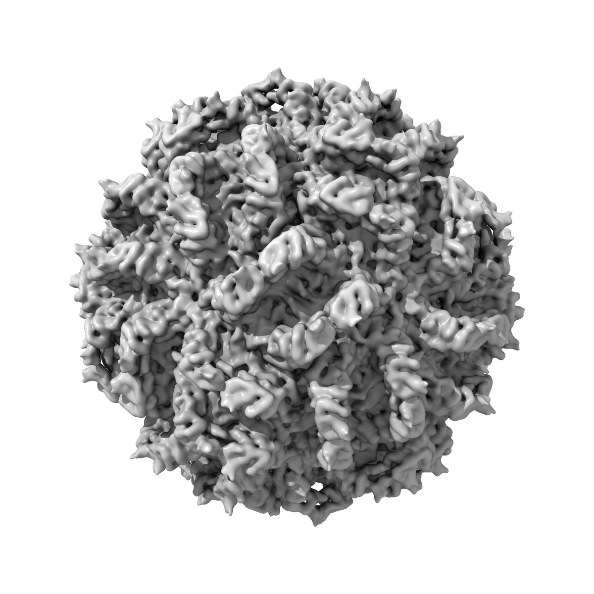
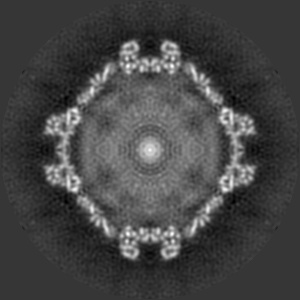
Partitivirus structure reveals a 120-subunit, helix-rich capsid with distinctive surface arches formed by quasisymmetric coat-protein dimers.
EMD-1459
Single-particle7.3 Å
 Deposition: 15/10/2007
Map released: 03/01/2008
Last modified: 11/12/2013
Sample Organism:
Penicillium stoloniferum virus S
Sample: PsV-S
Deposition Authors: Ochoa WF, Havens WM, Sinkovits RS ,
Nibert ML,
Ghabrial SA,
Baker TS
,
Nibert ML,
Ghabrial SA,
Baker TS
Sample: PsV-S
Deposition Authors: Ochoa WF, Havens WM, Sinkovits RS
 ,
Nibert ML,
Ghabrial SA,
Baker TS
,
Nibert ML,
Ghabrial SA,
Baker TS
Partitivirus structure reveals a 120-subunit, helix-rich capsid with distinctive surface arches formed by quasisymmetric coat-protein dimers.
Ochoa WF,
Havens WM,
Sinkovits RS ,
Nibert ML,
Ghabrial SA,
Baker TS
(2008) Structure , 16 , 776 - 786
,
Nibert ML,
Ghabrial SA,
Baker TS
(2008) Structure , 16 , 776 - 786
Abstract:
Two distinct partitiviruses, Penicillium stoloniferum viruses S and F, can be isolated from the fungus Penicillium stoloniferum. The bisegmented dsRNA genomes of these viruses are separately packaged in icosahedral capsids containing 120 coat-protein subunits. We used transmission electron cryomicroscopy and three-dimensional image reconstruction to determine the structure of Penicillium stoloniferum virus S at 7.3 A resolution. The capsid, approximately 350 A in outer diameter, contains 12 pentons, each of which is topped by five arched protrusions. Each of these protrusions is, in turn, formed by a quasisymmetric dimer of coat protein, for a total of 60 such dimers per particle. The density map shows numerous tubular features, characteristic of alpha helices and consistent with secondary structure predictions for the coat protein. This three-dimensional structure of a virus from the family Partitiviridae exhibits both similarities to and differences from the so-called "T = 2" capsids of other dsRNA viruses.
Two distinct partitiviruses, Penicillium stoloniferum viruses S and F, can be isolated from the fungus Penicillium stoloniferum. The bisegmented dsRNA genomes of these viruses are separately packaged in icosahedral capsids containing 120 coat-protein subunits. We used transmission electron cryomicroscopy and three-dimensional image reconstruction to determine the structure of Penicillium stoloniferum virus S at 7.3 A resolution. The capsid, approximately 350 A in outer diameter, contains 12 pentons, each of which is topped by five arched protrusions. Each of these protrusions is, in turn, formed by a quasisymmetric dimer of coat protein, for a total of 60 such dimers per particle. The density map shows numerous tubular features, characteristic of alpha helices and consistent with secondary structure predictions for the coat protein. This three-dimensional structure of a virus from the family Partitiviridae exhibits both similarities to and differences from the so-called "T = 2" capsids of other dsRNA viruses.