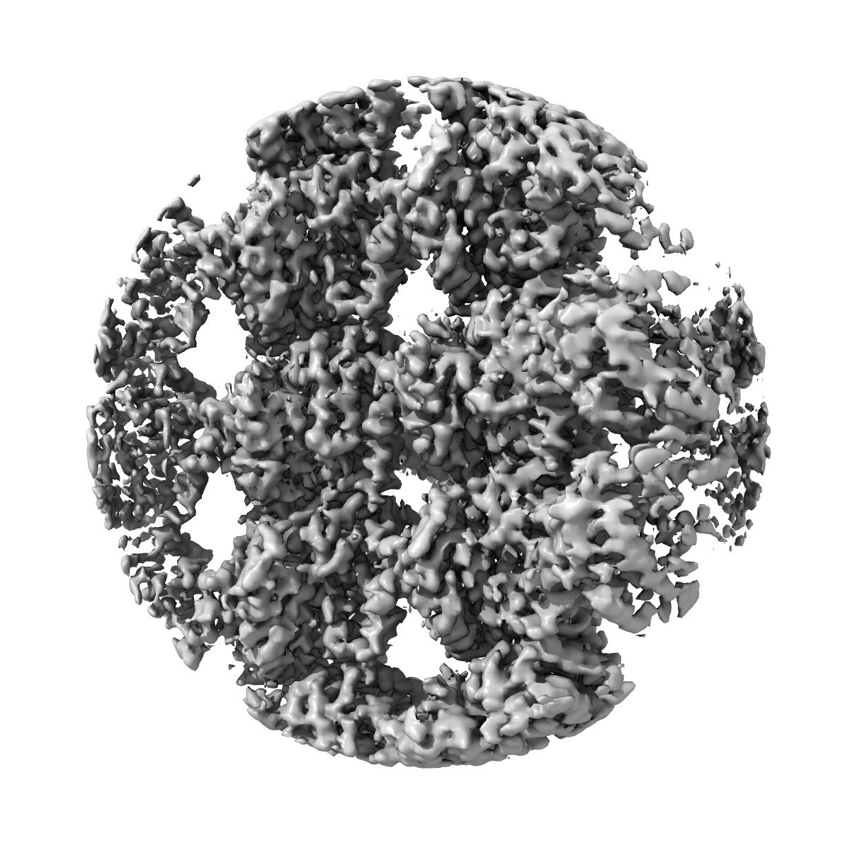
EMD-14634
Cryo-EM structure of GMPCPP-microtubules in complex with VASH2-SVBP
EMD-14634
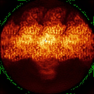
Single-particle3.6 Å
 Deposition: 29/03/2022
Map released: 14/12/2022
Last modified: 24/07/2024
Sample Organism:
Homo sapiens
Sample: VASH2-SVBP complex bound to the microtubule
Fitted models: 7zcw (Avg. Q-score: 0.509)
Deposition Authors: Choi SR ,
Blum T ,
Steinmetz MO
,
Blum T ,
Steinmetz MO
Sample: VASH2-SVBP complex bound to the microtubule
Fitted models: 7zcw (Avg. Q-score: 0.509)
Deposition Authors: Choi SR
 ,
Blum T ,
Steinmetz MO
,
Blum T ,
Steinmetz MO
VASH1-SVBP and VASH2-SVBP generate different detyrosination profiles on microtubules.
Ramirez-Rios S ,
Choi SR ,
Sanyal C ,
Blum TB ,
Bosc C ,
Krichen F ,
Denarier E ,
Soleilhac JM ,
Blot B ,
Janke C ,
Stoppin-Mellet V ,
Magiera MM ,
Arnal I ,
Steinmetz MO ,
Moutin MJ
(2023) J Cell Biol , 222
,
Choi SR ,
Sanyal C ,
Blum TB ,
Bosc C ,
Krichen F ,
Denarier E ,
Soleilhac JM ,
Blot B ,
Janke C ,
Stoppin-Mellet V ,
Magiera MM ,
Arnal I ,
Steinmetz MO ,
Moutin MJ
(2023) J Cell Biol , 222
Abstract:
The detyrosination/tyrosination cycle of α-tubulin is critical for proper cell functioning. VASH1-SVBP and VASH2-SVBP are ubiquitous enzymes involved in microtubule detyrosination, whose mode of action is little known. Here, we show in reconstituted systems and cells that VASH1-SVBP and VASH2-SVBP drive the global and local detyrosination of microtubules, respectively. We solved the cryo-electron microscopy structure of VASH2-SVBP bound to microtubules, revealing a different microtubule-binding configuration of its central catalytic region compared to VASH1-SVBP. We show that the divergent mode of detyrosination between the two enzymes is correlated with the microtubule-binding properties of their disordered N- and C-terminal regions. Specifically, the N-terminal region is responsible for a significantly longer residence time of VASH2-SVBP on microtubules compared to VASH1-SVBP. We suggest that this VASH region is critical for microtubule detachment and diffusion of VASH-SVBP enzymes on lattices. Our results suggest a mechanism by which VASH1-SVBP and VASH2-SVBP could generate distinct microtubule subpopulations and confined areas of detyrosinated lattices to drive various microtubule-based cellular functions.
The detyrosination/tyrosination cycle of α-tubulin is critical for proper cell functioning. VASH1-SVBP and VASH2-SVBP are ubiquitous enzymes involved in microtubule detyrosination, whose mode of action is little known. Here, we show in reconstituted systems and cells that VASH1-SVBP and VASH2-SVBP drive the global and local detyrosination of microtubules, respectively. We solved the cryo-electron microscopy structure of VASH2-SVBP bound to microtubules, revealing a different microtubule-binding configuration of its central catalytic region compared to VASH1-SVBP. We show that the divergent mode of detyrosination between the two enzymes is correlated with the microtubule-binding properties of their disordered N- and C-terminal regions. Specifically, the N-terminal region is responsible for a significantly longer residence time of VASH2-SVBP on microtubules compared to VASH1-SVBP. We suggest that this VASH region is critical for microtubule detachment and diffusion of VASH-SVBP enzymes on lattices. Our results suggest a mechanism by which VASH1-SVBP and VASH2-SVBP could generate distinct microtubule subpopulations and confined areas of detyrosinated lattices to drive various microtubule-based cellular functions.