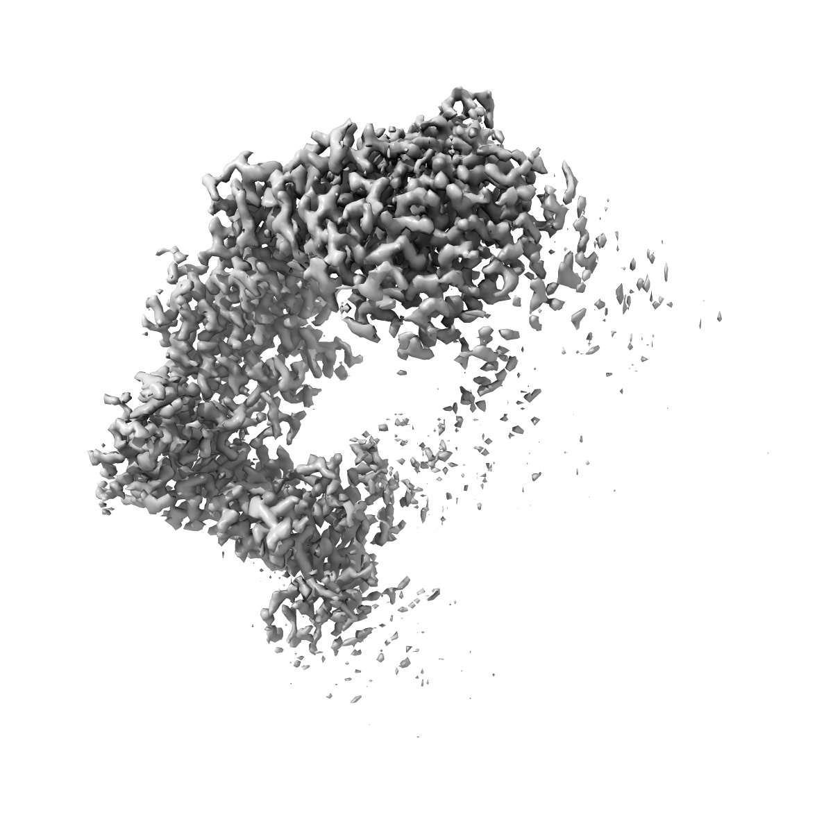
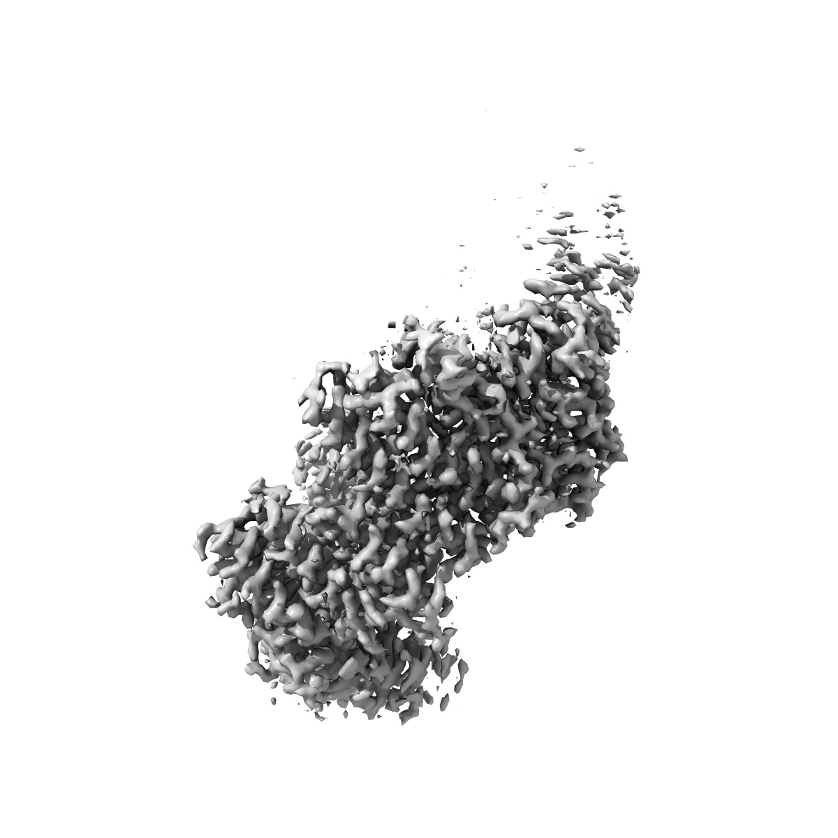
EMD-14719
FANCD2 Middle and C-terminal domains with USP1-NTE (focused refinement)
EMD-14719
Single-particle2.61 Å
 Deposition: 05/04/2022
Map released: 09/11/2022
Last modified: 13/12/2023
Sample Organism:
Homo sapiens
Sample: FANCD2 mono-ubiquitinated on K561
Raw data: EMPIAR-11299
Deposition Authors: Rennie ML ,
Walden H
,
Walden H
Sample: FANCD2 mono-ubiquitinated on K561
Raw data: EMPIAR-11299
Deposition Authors: Rennie ML
 ,
Walden H
,
Walden H
Cryo-EM reveals a mechanism of USP1 inhibition through a cryptic binding site.
Abstract:
Repair of DNA damage is critical to genomic integrity and frequently disrupted in cancers. Ubiquitin-specific protease 1 (USP1), a nucleus-localized deubiquitinase, lies at the interface of multiple DNA repair pathways and is a promising drug target for certain cancers. Although multiple inhibitors of this enzyme, including one in phase 1 clinical trials, have been established, their binding mode is unknown. Here, we use cryo-electron microscopy to study an assembled enzyme-substrate-inhibitor complex of USP1 and the well-established inhibitor, ML323. Achieving 2.5-Å resolution, with and without ML323, we find an unusual binding mode in which the inhibitor disrupts part of the hydrophobic core of USP1. The consequent conformational changes in the secondary structure lead to subtle rearrangements in the active site that underlie the mechanism of inhibition. These structures provide a platform for structure-based drug design targeting USP1.
Repair of DNA damage is critical to genomic integrity and frequently disrupted in cancers. Ubiquitin-specific protease 1 (USP1), a nucleus-localized deubiquitinase, lies at the interface of multiple DNA repair pathways and is a promising drug target for certain cancers. Although multiple inhibitors of this enzyme, including one in phase 1 clinical trials, have been established, their binding mode is unknown. Here, we use cryo-electron microscopy to study an assembled enzyme-substrate-inhibitor complex of USP1 and the well-established inhibitor, ML323. Achieving 2.5-Å resolution, with and without ML323, we find an unusual binding mode in which the inhibitor disrupts part of the hydrophobic core of USP1. The consequent conformational changes in the secondary structure lead to subtle rearrangements in the active site that underlie the mechanism of inhibition. These structures provide a platform for structure-based drug design targeting USP1.