EMD-15754
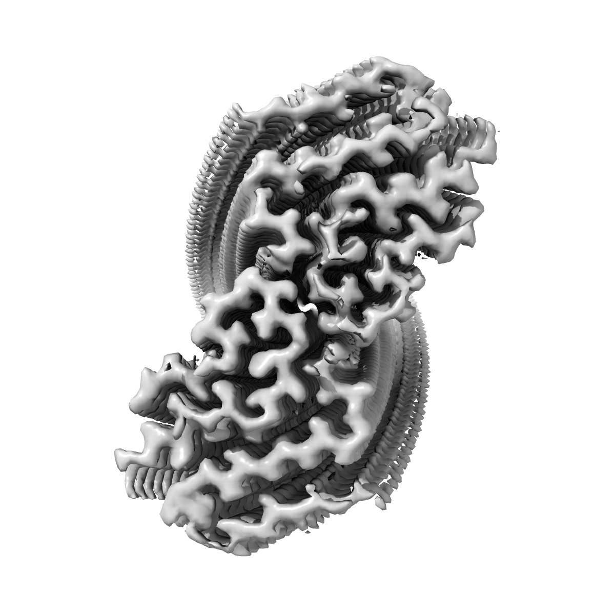
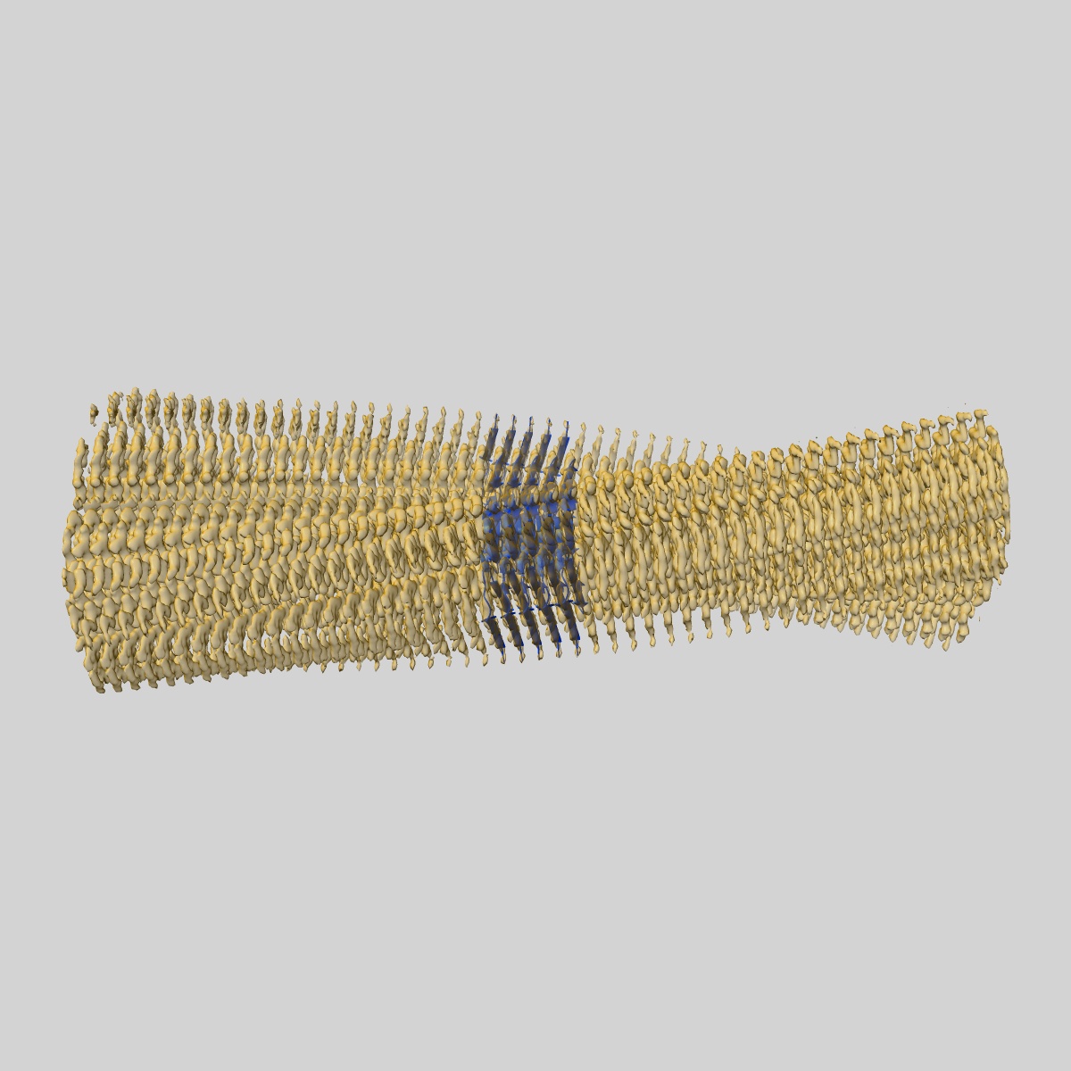
IAPP S20G plateau-phase fibril polymorph 4PF-CU
EMD-15754
Helical reconstruction2.3 Å
 Deposition: 05/09/2022
Map released: 10/01/2024
Last modified: 23/10/2024
Sample Organism:
Homo sapiens
Sample: IAPP S20G plateau-phase fibril polymorph 4PF-CU
Fitted models: 8az5 (Avg. Q-score: 0.638)
Raw data: EMPIAR-11717
Deposition Authors: Wilkinson M, Xu Y, Gallardo R, Radford SE, Ranson NA
Sample: IAPP S20G plateau-phase fibril polymorph 4PF-CU
Fitted models: 8az5 (Avg. Q-score: 0.638)
Raw data: EMPIAR-11717
Deposition Authors: Wilkinson M, Xu Y, Gallardo R, Radford SE, Ranson NA

Structural evolution of fibril polymorphs during amyloid assembly.
Wilkinson M,
Xu Y,
Thacker D,
Taylor AIP ,
Fisher DG,
Gallardo RU,
Radford SE,
Ranson NA
(2023) Cell , 186 , 5798 - 5811.e26
,
Fisher DG,
Gallardo RU,
Radford SE,
Ranson NA
(2023) Cell , 186 , 5798 - 5811.e26
Abstract:
Cryoelectron microscopy (cryo-EM) has provided unprecedented insights into amyloid fibril structures, including those associated with disease. However, these structures represent the endpoints of long assembly processes, and their relationship to fibrils formed early in assembly is unknown. Consequently, whether different fibril architectures, with potentially different pathological properties, form during assembly remains unknown. Here, we used cryo-EM to determine structures of amyloid fibrils at different times during in vitro fibrillation of a disease-related variant of human islet amyloid polypeptide (IAPP-S20G). Strikingly, the fibrils formed in the lag, growth, and plateau phases have different structures, with new forms appearing and others disappearing as fibrillation proceeds. A time course with wild-type hIAPP also shows fibrils changing with time, suggesting that this is a general property of IAPP amyloid assembly. The observation of transiently populated fibril structures has implications for understanding amyloid assembly mechanisms with potential new insights into amyloid progression in disease.
Cryoelectron microscopy (cryo-EM) has provided unprecedented insights into amyloid fibril structures, including those associated with disease. However, these structures represent the endpoints of long assembly processes, and their relationship to fibrils formed early in assembly is unknown. Consequently, whether different fibril architectures, with potentially different pathological properties, form during assembly remains unknown. Here, we used cryo-EM to determine structures of amyloid fibrils at different times during in vitro fibrillation of a disease-related variant of human islet amyloid polypeptide (IAPP-S20G). Strikingly, the fibrils formed in the lag, growth, and plateau phases have different structures, with new forms appearing and others disappearing as fibrillation proceeds. A time course with wild-type hIAPP also shows fibrils changing with time, suggesting that this is a general property of IAPP amyloid assembly. The observation of transiently populated fibril structures has implications for understanding amyloid assembly mechanisms with potential new insights into amyloid progression in disease.