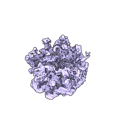
EMD-16498
Cryo-EM captures early ribosome assembly in action
EMD-16498
Single-particle4.19 Å
 Deposition: 21/01/2023
Map released: 05/04/2023
Last modified: 24/07/2024
Sample Organism:
Escherichia coli
Sample: large ribosomal subunit precursor C_L2
Fitted models: 8c91 (Avg. Q-score: 0.276)
Deposition Authors: Lauer S, Nikolay R ,
Qin B
Sample: large ribosomal subunit precursor C_L2
Fitted models: 8c91 (Avg. Q-score: 0.276)
Deposition Authors: Lauer S, Nikolay R
 ,
Qin B
Cryo-EM captures early ribosome assembly in action.
Qin B  ,
Lauer SM
  ,
Balke A
  ,
Vieira-Vieira CH
  ,
Burger J
  ,
Mielke T
  ,
Selbach M
  ,
Scheerer P
  ,
Spahn CMT
  ,
Nikolay R
(2023) Nat Commun , 14 , 898 - 898
 ,
Lauer SM
  ,
Balke A
  ,
Vieira-Vieira CH
  ,
Burger J
  ,
Mielke T
  ,
Selbach M
  ,
Scheerer P
  ,
Spahn CMT
  ,
Nikolay R
(2023) Nat Commun , 14 , 898 - 898
Abstract:
Ribosome biogenesis is a fundamental multi-step cellular process in all domains of life that involves the production, processing, folding, and modification of ribosomal RNAs (rRNAs) and ribosomal proteins. To obtain insights into the still unexplored early assembly phase of the bacterial 50S subunit, we exploited a minimal in vitro reconstitution system using purified ribosomal components and scalable reaction conditions. Time-limited assembly assays combined with cryo-EM analysis visualizes the structurally complex assembly pathway starting with a particle consisting of ordered density for only ~500 nucleotides of 23S rRNA domain I and three ribosomal proteins. In addition, our structural analysis reveals that early 50S assembly occurs in a domain-wise fashion, while late 50S assembly proceeds incrementally. Furthermore, we find that both ribosomal proteins and folded rRNA helices, occupying surface exposed regions on pre-50S particles, induce, or stabilize rRNA folds within adjacent regions, thereby creating cooperativity.
Ribosome biogenesis is a fundamental multi-step cellular process in all domains of life that involves the production, processing, folding, and modification of ribosomal RNAs (rRNAs) and ribosomal proteins. To obtain insights into the still unexplored early assembly phase of the bacterial 50S subunit, we exploited a minimal in vitro reconstitution system using purified ribosomal components and scalable reaction conditions. Time-limited assembly assays combined with cryo-EM analysis visualizes the structurally complex assembly pathway starting with a particle consisting of ordered density for only ~500 nucleotides of 23S rRNA domain I and three ribosomal proteins. In addition, our structural analysis reveals that early 50S assembly occurs in a domain-wise fashion, while late 50S assembly proceeds incrementally. Furthermore, we find that both ribosomal proteins and folded rRNA helices, occupying surface exposed regions on pre-50S particles, induce, or stabilize rRNA folds within adjacent regions, thereby creating cooperativity.
