EMD-1756
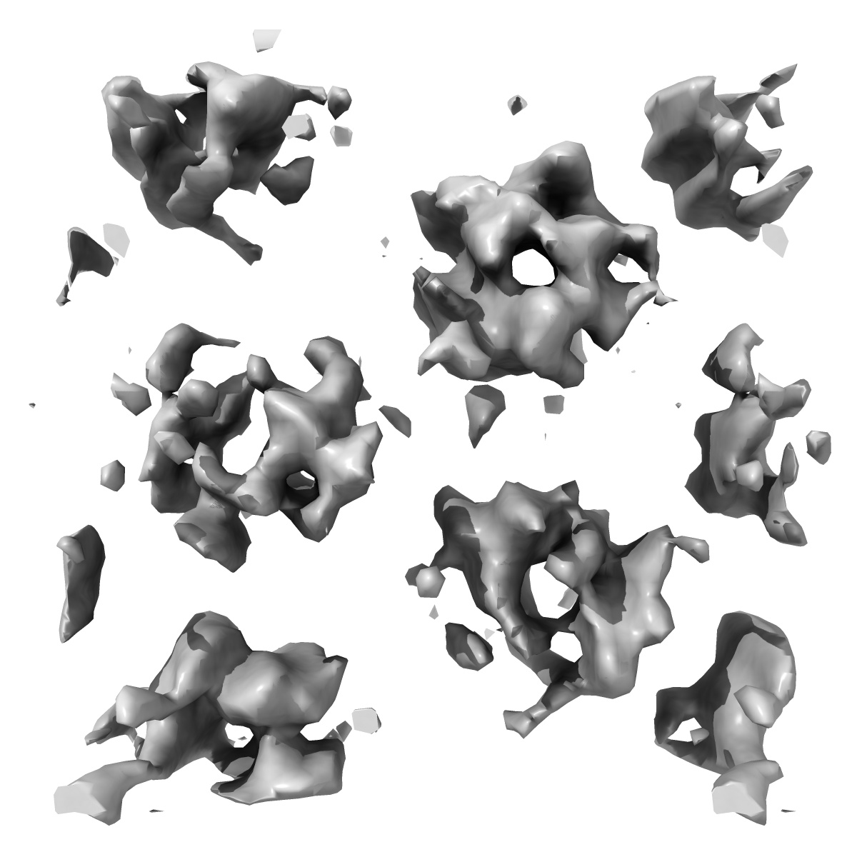
Electron crystallography reveals the asymmetry of the trimeric glycine betaine transporter BetP from Corynebacterium glutamicum
EMD-1756
Electron Crystallography8.0 Å
 Deposition: 07/07/2010
Map released: 23/07/2010
Last modified: 24/02/2012
Sample Organism:
Corynebacterium glutamicum
Sample: Sodium/betaine symporter BetP with 45 amino acids truncated at the C-terminus
Deposition Authors: Tsai C-J, Khafizov KF, Hakulinen J, Forrest LR, Kraemer R, Kuehlbrandt W, Ziegler C
Sample: Sodium/betaine symporter BetP with 45 amino acids truncated at the C-terminus
Deposition Authors: Tsai C-J, Khafizov KF, Hakulinen J, Forrest LR, Kraemer R, Kuehlbrandt W, Ziegler C
Structural asymmetry in a trimeric Na+/betaine symporter, BetP, from Corynebacterium glutamicum.
Tsai C-J  ,
Khafizov KF,
Hakulinen J,
Forrest LR,
Kraemer R,
Kuehlbrandt W,
Ziegler C
,
Khafizov KF,
Hakulinen J,
Forrest LR,
Kraemer R,
Kuehlbrandt W,
Ziegler C
(2011) J. Mol. Biol. , 407 , 368 - 381
 ,
Khafizov KF,
Hakulinen J,
Forrest LR,
Kraemer R,
Kuehlbrandt W,
Ziegler C
,
Khafizov KF,
Hakulinen J,
Forrest LR,
Kraemer R,
Kuehlbrandt W,
Ziegler C
(2011) J. Mol. Biol. , 407 , 368 - 381
Abstract:
The Na(+)-coupled symporter BetP catalyzes the uptake of the compatible solute betaine in the soil bacterium Corynebacterium glutamicum. BetP also senses hyperosmotic stress and regulates its own activity in response to stress level. We determined a three-dimensional (3D) map (at 8 Å in-plane resolution) of a constitutively active mutant of BetP in a C. glutamicum membrane environment by electron cryomicroscopy of two-dimensional crystals. The map shows that the constitutively active mutant, which lacks the C-terminal domain involved in osmosensing, is trimeric like wild-type BetP. Recently, we reported the X-ray crystal structure of BetP at 3.35 Å, in which all three protomers displayed a substrate-occluded state. Rigid-body fitting of this trimeric structure to the 3D map identified the periplasmic and cytoplasmic sides of the membrane. Fitting of an X-ray monomer to the individual protomer maps allowed assignment of transmembrane helices and of the substrate pathway, and revealed differences in trimer architecture from the X-ray structure in the tilt angle of each protomer with respect to the membrane. The three protomer maps showed pronounced differences around the substrate pathway, suggesting three different conformations within the same trimer. Two of those protomer maps closely match those of the atomic structures of the outward-facing and inward-facing states of the hydantoin transporter Mhp1, suggesting that the BetP protomer conformations reflect key states of the transport cycle. Thus, the asymmetry in the two-dimensional maps may reflect cooperativity of conformational changes within the BetP trimer, which potentially increases the rate of glycine betaine uptake.
The Na(+)-coupled symporter BetP catalyzes the uptake of the compatible solute betaine in the soil bacterium Corynebacterium glutamicum. BetP also senses hyperosmotic stress and regulates its own activity in response to stress level. We determined a three-dimensional (3D) map (at 8 Å in-plane resolution) of a constitutively active mutant of BetP in a C. glutamicum membrane environment by electron cryomicroscopy of two-dimensional crystals. The map shows that the constitutively active mutant, which lacks the C-terminal domain involved in osmosensing, is trimeric like wild-type BetP. Recently, we reported the X-ray crystal structure of BetP at 3.35 Å, in which all three protomers displayed a substrate-occluded state. Rigid-body fitting of this trimeric structure to the 3D map identified the periplasmic and cytoplasmic sides of the membrane. Fitting of an X-ray monomer to the individual protomer maps allowed assignment of transmembrane helices and of the substrate pathway, and revealed differences in trimer architecture from the X-ray structure in the tilt angle of each protomer with respect to the membrane. The three protomer maps showed pronounced differences around the substrate pathway, suggesting three different conformations within the same trimer. Two of those protomer maps closely match those of the atomic structures of the outward-facing and inward-facing states of the hydantoin transporter Mhp1, suggesting that the BetP protomer conformations reflect key states of the transport cycle. Thus, the asymmetry in the two-dimensional maps may reflect cooperativity of conformational changes within the BetP trimer, which potentially increases the rate of glycine betaine uptake.