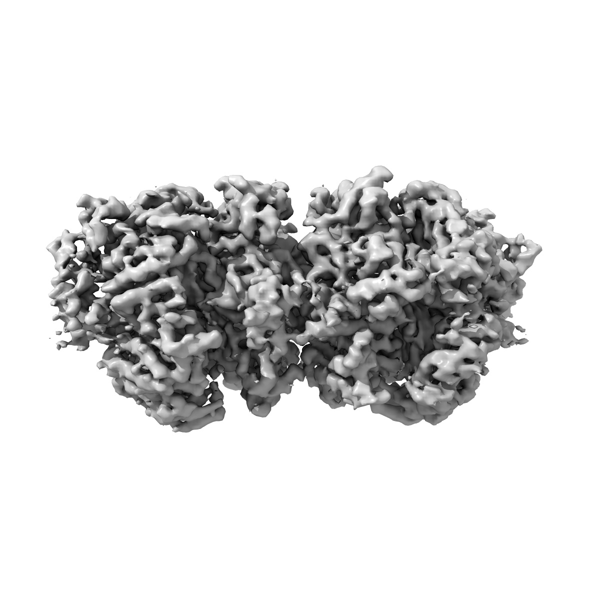
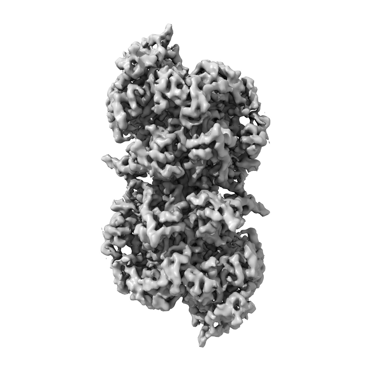
EMD-19625
H. sapiens MCM double hexamer containing the MCM5 R195A/L209G mutant
EMD-19625
Single-particle5.6 Å
 Deposition: 13/02/2024
Map released: 02/10/2024
Last modified: 25/12/2024
Sample Organism:
Homo sapiens
Sample: H. sapiens MCM double hexamer containing the MCM5 R195A/L209G mutant
Deposition Authors: Puehringer T ,
Weissmann F ,
Greiwe J ,
Diffley JFX ,
Costa A
,
Weissmann F ,
Greiwe J ,
Diffley JFX ,
Costa A
Sample: H. sapiens MCM double hexamer containing the MCM5 R195A/L209G mutant
Deposition Authors: Puehringer T
 ,
Weissmann F ,
Greiwe J ,
Diffley JFX ,
Costa A
,
Weissmann F ,
Greiwe J ,
Diffley JFX ,
Costa A
MCM double hexamer loading visualized with human proteins.
Weissmann F ,
Greiwe JF ,
Puhringer T ,
Eastwood EL ,
Couves EC ,
Miller TCR ,
Diffley JFX ,
Costa A
(2024) Nature , 636 , 499 - 508
,
Greiwe JF ,
Puhringer T ,
Eastwood EL ,
Couves EC ,
Miller TCR ,
Diffley JFX ,
Costa A
(2024) Nature , 636 , 499 - 508
Abstract:
Eukaryotic DNA replication begins with the loading of the MCM replicative DNA helicase as a head-to-head double hexamer at origins of DNA replication1-3. Our current understanding of how the double hexamer is assembled by the origin recognition complex (ORC), CDC6 and CDT1 comes mostly from budding yeast. Here we characterize human double hexamer (hDH) loading using biochemical reconstitution and cryo-electron microscopy with purified proteins. We show that the human double hexamer engages DNA differently from the yeast double hexamer (yDH), and generates approximately five base pairs of underwound DNA at the interface between hexamers, as seen in hDH isolated from cells4. We identify several differences from the yeast double hexamer in the order of factor recruitment and dependencies during hDH assembly. Unlike in yeast5-8, the ORC6 subunit of the ORC is not essential for initial MCM recruitment or hDH loading, but contributes to an alternative hDH assembly pathway that requires an intrinsically disordered region in ORC1, which may work through a MCM-ORC intermediate. Our work presents a detailed view of how double hexamers are assembled in an organism that uses sequence-independent replication origins, provides further evidence for diversity in eukaryotic double hexamer assembly mechanisms9, and represents a first step towards reconstitution of DNA replication initiation with purified human proteins.
Eukaryotic DNA replication begins with the loading of the MCM replicative DNA helicase as a head-to-head double hexamer at origins of DNA replication1-3. Our current understanding of how the double hexamer is assembled by the origin recognition complex (ORC), CDC6 and CDT1 comes mostly from budding yeast. Here we characterize human double hexamer (hDH) loading using biochemical reconstitution and cryo-electron microscopy with purified proteins. We show that the human double hexamer engages DNA differently from the yeast double hexamer (yDH), and generates approximately five base pairs of underwound DNA at the interface between hexamers, as seen in hDH isolated from cells4. We identify several differences from the yeast double hexamer in the order of factor recruitment and dependencies during hDH assembly. Unlike in yeast5-8, the ORC6 subunit of the ORC is not essential for initial MCM recruitment or hDH loading, but contributes to an alternative hDH assembly pathway that requires an intrinsically disordered region in ORC1, which may work through a MCM-ORC intermediate. Our work presents a detailed view of how double hexamers are assembled in an organism that uses sequence-independent replication origins, provides further evidence for diversity in eukaryotic double hexamer assembly mechanisms9, and represents a first step towards reconstitution of DNA replication initiation with purified human proteins.
Secondary citations:
- Weissmann F, Greiwe JF, Puhringer T, Miller TCR, Diffley JFX & Costa A. (2024) MCM Double Hexamer Loading Visualised with Human Proteins. bioRxiv,
- Liebschner D, Afonine PV, Baker ML, Bunkoczi G, Chen VB, Croll TI, Hintze B, Hung LW, Jain S, McCoy AJ, Moriarty NW, Oeffner RD, Poon BK, Prisant MG, Read RJ, Richardson JS, Richardson DC, Sammito MD, Sobolev OV, Stockwell DH, Terwilliger TC, Urzhumtsev AG, Videau LL, Williams CJ & Adams PD. (2019) Macromolecular structure determination using X-rays, neutrons and electrons: recent developments in Phenix. Acta Crystallogr D Struct Biol, 75, 861 - 877