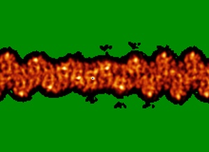
EMD-1990
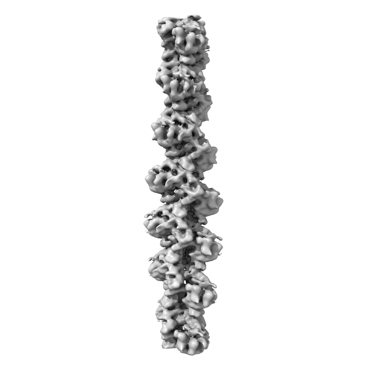
Structure of bare F-actin filaments obtained from the same sample as the Actin-Tropomyosin-Myosin Complex
EMD-1990
Helical reconstruction8.9 Å
 Deposition: 14/11/2011
Map released: 01/08/2012
Last modified: 01/08/2012
Sample Organism:
Oryctolagus cuniculus
Sample: Bare F-actin filaments
Fitted models: 4a7n (Avg. Q-score: 0.069)
Deposition Authors: Behrmann E, Mueller M, Penczek PA, Mannherz HG, Manstein DJ, Raunser S
Sample: Bare F-actin filaments
Fitted models: 4a7n (Avg. Q-score: 0.069)
Deposition Authors: Behrmann E, Mueller M, Penczek PA, Mannherz HG, Manstein DJ, Raunser S
Structure of the rigor actin-tropomyosin-myosin complex.
Behrmann E  ,
Mueller M,
Penczek PA,
Mannherz HG ,
Manstein DJ ,
Raunser S
,
Mueller M,
Penczek PA,
Mannherz HG ,
Manstein DJ ,
Raunser S
(2012) Cell , 150 , 327 - 338
 ,
Mueller M,
Penczek PA,
Mannherz HG ,
Manstein DJ ,
Raunser S
,
Mueller M,
Penczek PA,
Mannherz HG ,
Manstein DJ ,
Raunser S
(2012) Cell , 150 , 327 - 338
Abstract:
Regulation of myosin and filamentous actin interaction by tropomyosin is a central feature of contractile events in muscle and nonmuscle cells. However, little is known about molecular interactions within the complex and the trajectory of tropomyosin movement between its "open" and "closed" positions on the actin filament. Here, we report the 8 Å resolution structure of the rigor (nucleotide-free) actin-tropomyosin-myosin complex determined by cryo-electron microscopy. The pseudoatomic model of the complex, obtained from fitting crystal structures into the map, defines the large interface involving two adjacent actin monomers and one tropomyosin pseudorepeat per myosin contact. Severe forms of hereditary myopathies are linked to mutations that critically perturb this interface. Myosin binding results in a 23 Å shift of tropomyosin along actin. Complex domain motions occur in myosin, but not in actin. Based on our results, we propose a structural model for the tropomyosin-dependent modulation of myosin binding to actin.
Regulation of myosin and filamentous actin interaction by tropomyosin is a central feature of contractile events in muscle and nonmuscle cells. However, little is known about molecular interactions within the complex and the trajectory of tropomyosin movement between its "open" and "closed" positions on the actin filament. Here, we report the 8 Å resolution structure of the rigor (nucleotide-free) actin-tropomyosin-myosin complex determined by cryo-electron microscopy. The pseudoatomic model of the complex, obtained from fitting crystal structures into the map, defines the large interface involving two adjacent actin monomers and one tropomyosin pseudorepeat per myosin contact. Severe forms of hereditary myopathies are linked to mutations that critically perturb this interface. Myosin binding results in a 23 Å shift of tropomyosin along actin. Complex domain motions occur in myosin, but not in actin. Based on our results, we propose a structural model for the tropomyosin-dependent modulation of myosin binding to actin.