EMD-20051
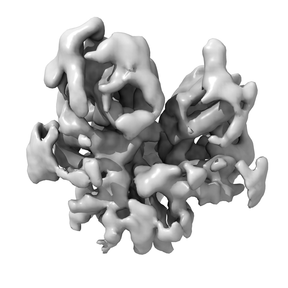
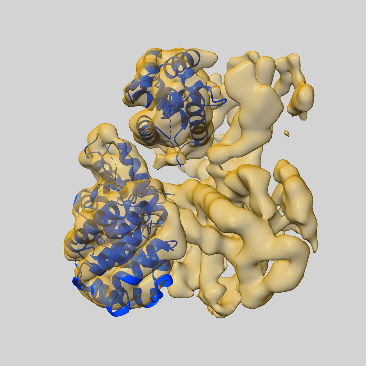
CryoEM focus classification map of the hyperactive ClpB mutant K476C, bound to casein, NTD-trimer
EMD-20051
Single-particle4.1 Å
 Deposition: 01/04/2019
Map released: 12/06/2019
Last modified: 20/03/2024
Sample Organism:
Escherichia coli K-12,
Bos taurus
Sample: hyperactive ClpB mutant K476C bound to casein
Fitted models: 6og3 (Avg. Q-score: 0.306)
Deposition Authors: Rizo AR, Lin J-B
Sample: hyperactive ClpB mutant K476C bound to casein
Fitted models: 6og3 (Avg. Q-score: 0.306)
Deposition Authors: Rizo AR, Lin J-B
Structural basis for substrate gripping and translocation by the ClpB AAA+ disaggregase.
Rizo AN,
Lin J,
Gates SN,
Tse E  ,
Bart SM,
Castellano LM,
DiMaio F,
Shorter J ,
Southworth DR
,
Bart SM,
Castellano LM,
DiMaio F,
Shorter J ,
Southworth DR
(2019) Nat Commun , 10 , 2393 - 2393
 ,
Bart SM,
Castellano LM,
DiMaio F,
Shorter J ,
Southworth DR
,
Bart SM,
Castellano LM,
DiMaio F,
Shorter J ,
Southworth DR
(2019) Nat Commun , 10 , 2393 - 2393
Abstract:
Bacterial ClpB and yeast Hsp104 are homologous Hsp100 protein disaggregases that serve critical functions in proteostasis by solubilizing protein aggregates. Two AAA+ nucleotide binding domains (NBDs) power polypeptide translocation through a central channel comprised of a hexameric spiral of protomers that contact substrate via conserved pore-loop interactions. Here we report cryo-EM structures of a hyperactive ClpB variant bound to the model substrate, casein in the presence of slowly hydrolysable ATPγS, which reveal the translocation mechanism. Distinct substrate-gripping interactions are identified for NBD1 and NBD2 pore loops. A trimer of N-terminal domains define a channel entrance that binds the polypeptide substrate adjacent to the topmost NBD1 contact. NBD conformations at the seam interface reveal how ATP hydrolysis-driven substrate disengagement and re-binding are precisely tuned to drive a directional, stepwise translocation cycle.
Bacterial ClpB and yeast Hsp104 are homologous Hsp100 protein disaggregases that serve critical functions in proteostasis by solubilizing protein aggregates. Two AAA+ nucleotide binding domains (NBDs) power polypeptide translocation through a central channel comprised of a hexameric spiral of protomers that contact substrate via conserved pore-loop interactions. Here we report cryo-EM structures of a hyperactive ClpB variant bound to the model substrate, casein in the presence of slowly hydrolysable ATPγS, which reveal the translocation mechanism. Distinct substrate-gripping interactions are identified for NBD1 and NBD2 pore loops. A trimer of N-terminal domains define a channel entrance that binds the polypeptide substrate adjacent to the topmost NBD1 contact. NBD conformations at the seam interface reveal how ATP hydrolysis-driven substrate disengagement and re-binding are precisely tuned to drive a directional, stepwise translocation cycle.