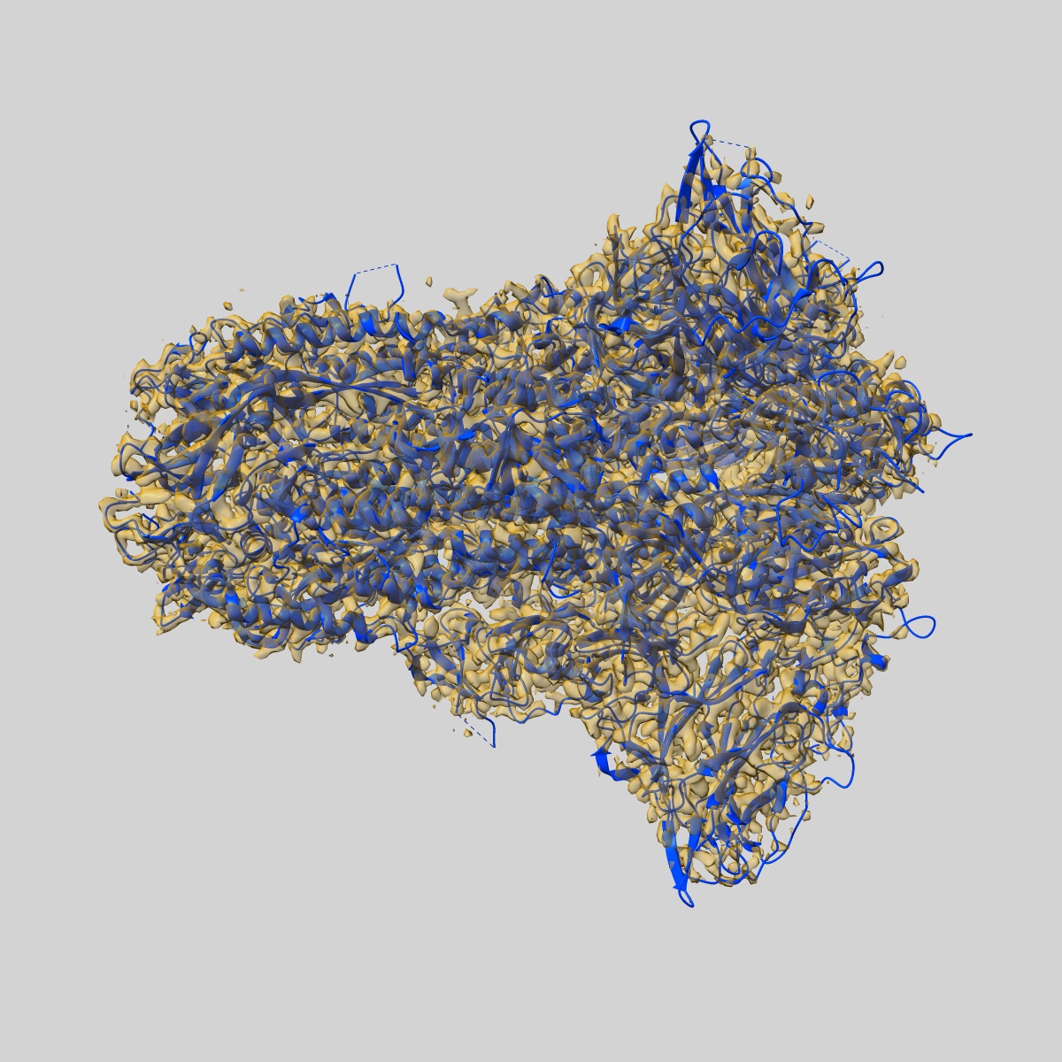
EMD-20070
Structural basis for human coronavirus attachment to sialic acid receptors. Apo-HCoV-OC43 S
EMD-20070
Single-particle2.9 Å
 Deposition: 07/04/2019
Map released: 05/06/2019
Last modified: 06/11/2024
Sample Organism:
Betacoronavirus,
Human coronavirus OC43
Sample: HCoV-OC43 spike glycoprotein ectodomain in complex with 9-O-acetyl sialic acid
Fitted models: 6ohw (Avg. Q-score: 0.561)
Deposition Authors: Tortorici MA, Walls AC
Sample: HCoV-OC43 spike glycoprotein ectodomain in complex with 9-O-acetyl sialic acid
Fitted models: 6ohw (Avg. Q-score: 0.561)
Deposition Authors: Tortorici MA, Walls AC

Structural basis for human coronavirus attachment to sialic acid receptors.
Alejandra Tortorici M ,
Walls AC ,
Lang Y,
Wang C ,
Li Z ,
Koerhuis D,
Boons GJ ,
Bosch BJ,
Rey FA ,
de Groot RJ,
Veesler D
(2019) Nat Struct Mol Biol , 26 , 481 - 489
,
Walls AC ,
Lang Y,
Wang C ,
Li Z ,
Koerhuis D,
Boons GJ ,
Bosch BJ,
Rey FA ,
de Groot RJ,
Veesler D
(2019) Nat Struct Mol Biol , 26 , 481 - 489
Abstract:
Coronaviruses cause respiratory tract infections in humans and outbreaks of deadly pneumonia worldwide. Infections are initiated by the transmembrane spike (S) glycoprotein, which binds to host receptors and fuses the viral and cellular membranes. To understand the molecular basis of coronavirus attachment to oligosaccharide receptors, we determined cryo-EM structures of coronavirus OC43 S glycoprotein trimer in isolation and in complex with a 9-O-acetylated sialic acid. We show that the ligand binds with fast kinetics to a surface-exposed groove and that interactions at the identified site are essential for S-mediated viral entry into host cells, but free monosaccharide does not trigger fusogenic conformational changes. The receptor-interacting site is conserved in all coronavirus S glycoproteins that engage 9-O-acetyl-sialogycans, with an architecture similar to those of the ligand-binding pockets of coronavirus hemagglutinin esterases and influenza virus C/D hemagglutinin-esterase fusion glycoproteins. Our results demonstrate these viruses evolved similar strategies to engage sialoglycans at the surface of target cells.
Coronaviruses cause respiratory tract infections in humans and outbreaks of deadly pneumonia worldwide. Infections are initiated by the transmembrane spike (S) glycoprotein, which binds to host receptors and fuses the viral and cellular membranes. To understand the molecular basis of coronavirus attachment to oligosaccharide receptors, we determined cryo-EM structures of coronavirus OC43 S glycoprotein trimer in isolation and in complex with a 9-O-acetylated sialic acid. We show that the ligand binds with fast kinetics to a surface-exposed groove and that interactions at the identified site are essential for S-mediated viral entry into host cells, but free monosaccharide does not trigger fusogenic conformational changes. The receptor-interacting site is conserved in all coronavirus S glycoproteins that engage 9-O-acetyl-sialogycans, with an architecture similar to those of the ligand-binding pockets of coronavirus hemagglutinin esterases and influenza virus C/D hemagglutinin-esterase fusion glycoproteins. Our results demonstrate these viruses evolved similar strategies to engage sialoglycans at the surface of target cells.