EMD-20162
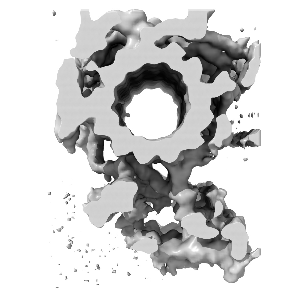
The central pair apparatus focusing on the C1a-e-c supercomplex extracted from the cryo-electron tomography and subtomographic average of isolated Chlamydomonas fap76-1 mutant axoneme
EMD-20162
Subtomogram averaging22.0 Å
 Deposition: 26/04/2019
Map released: 13/11/2019
Last modified: 12/08/2020
Sample Organism:
Chlamydomonas reinhardtii
Sample: The C1a-e-c supercomplex of central pair apparatus averaged from Chlamydomonas fap76-1 mutant cilia
Deposition Authors: Fu G, Nicastro D
Sample: The C1a-e-c supercomplex of central pair apparatus averaged from Chlamydomonas fap76-1 mutant cilia
Deposition Authors: Fu G, Nicastro D
Structural organization of the C1a-e-c supercomplex within the ciliary central apparatus.
Fu G  ,
Zhao L,
Dymek E,
Hou Y ,
Song K,
Phan N,
Shang Z,
Smith EF,
Witman GB ,
Nicastro D
,
Zhao L,
Dymek E,
Hou Y ,
Song K,
Phan N,
Shang Z,
Smith EF,
Witman GB ,
Nicastro D
(2019) J. Cell Biol. , 218 , 4236 - 4251
 ,
Zhao L,
Dymek E,
Hou Y ,
Song K,
Phan N,
Shang Z,
Smith EF,
Witman GB ,
Nicastro D
,
Zhao L,
Dymek E,
Hou Y ,
Song K,
Phan N,
Shang Z,
Smith EF,
Witman GB ,
Nicastro D
(2019) J. Cell Biol. , 218 , 4236 - 4251
Abstract:
Nearly all motile cilia contain a central apparatus (CA) composed of two connected singlet microtubules with attached projections that play crucial roles in regulating ciliary motility. Defects in CA assembly usually result in motility-impaired or paralyzed cilia, which in humans causes disease. Despite their importance, the protein composition and functions of the CA projections are largely unknown. Here, we integrated biochemical and genetic approaches with cryo-electron tomography to compare the CA of wild-type Chlamydomonas with CA mutants. We identified a large (>2 MD) complex, the C1a-e-c supercomplex, that requires the PF16 protein for assembly and contains the CA components FAP76, FAP81, FAP92, and FAP216. We localized these subunits within the supercomplex using nanogold labeling and show that loss of any one of them results in impaired ciliary motility. These data provide insight into the subunit organization and 3D structure of the CA, which is a prerequisite for understanding the molecular mechanisms by which the CA regulates ciliary beating.
Nearly all motile cilia contain a central apparatus (CA) composed of two connected singlet microtubules with attached projections that play crucial roles in regulating ciliary motility. Defects in CA assembly usually result in motility-impaired or paralyzed cilia, which in humans causes disease. Despite their importance, the protein composition and functions of the CA projections are largely unknown. Here, we integrated biochemical and genetic approaches with cryo-electron tomography to compare the CA of wild-type Chlamydomonas with CA mutants. We identified a large (>2 MD) complex, the C1a-e-c supercomplex, that requires the PF16 protein for assembly and contains the CA components FAP76, FAP81, FAP92, and FAP216. We localized these subunits within the supercomplex using nanogold labeling and show that loss of any one of them results in impaired ciliary motility. These data provide insight into the subunit organization and 3D structure of the CA, which is a prerequisite for understanding the molecular mechanisms by which the CA regulates ciliary beating.