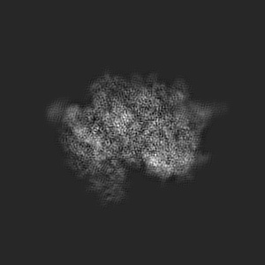
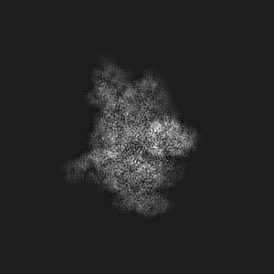
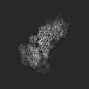
EMD-20249
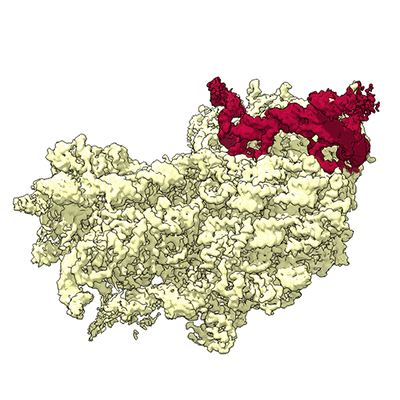
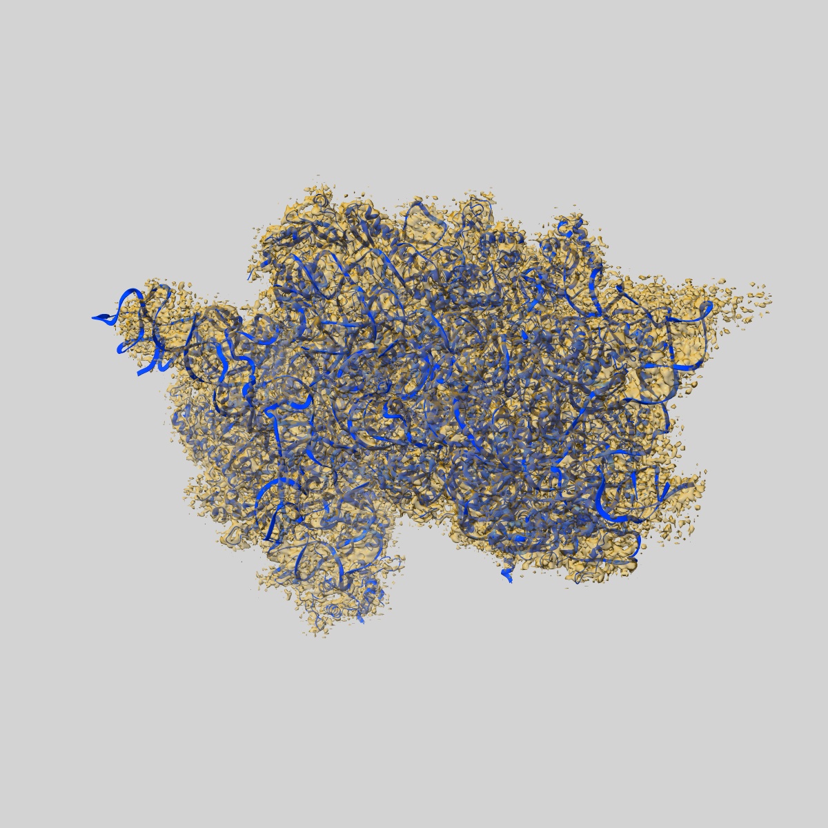
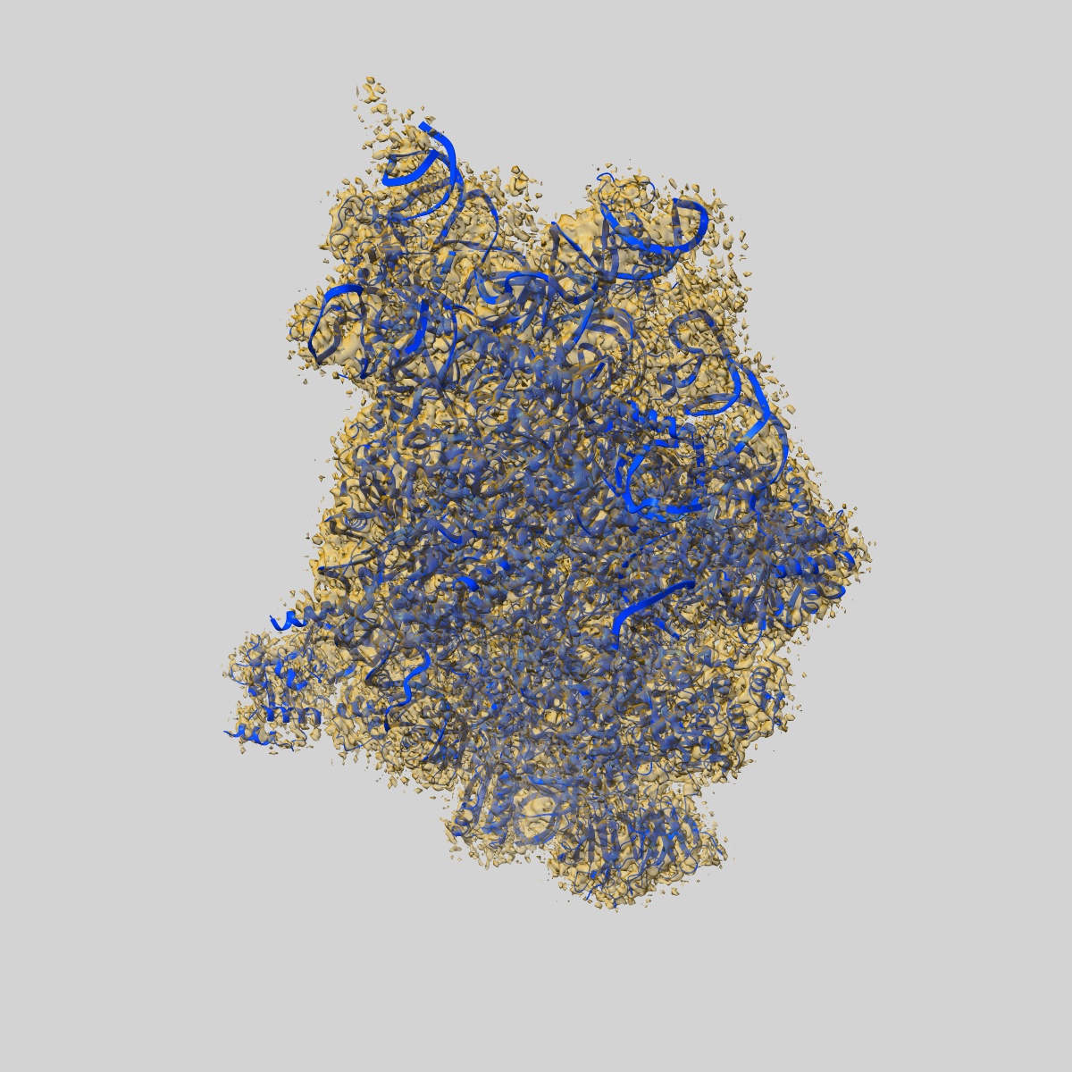
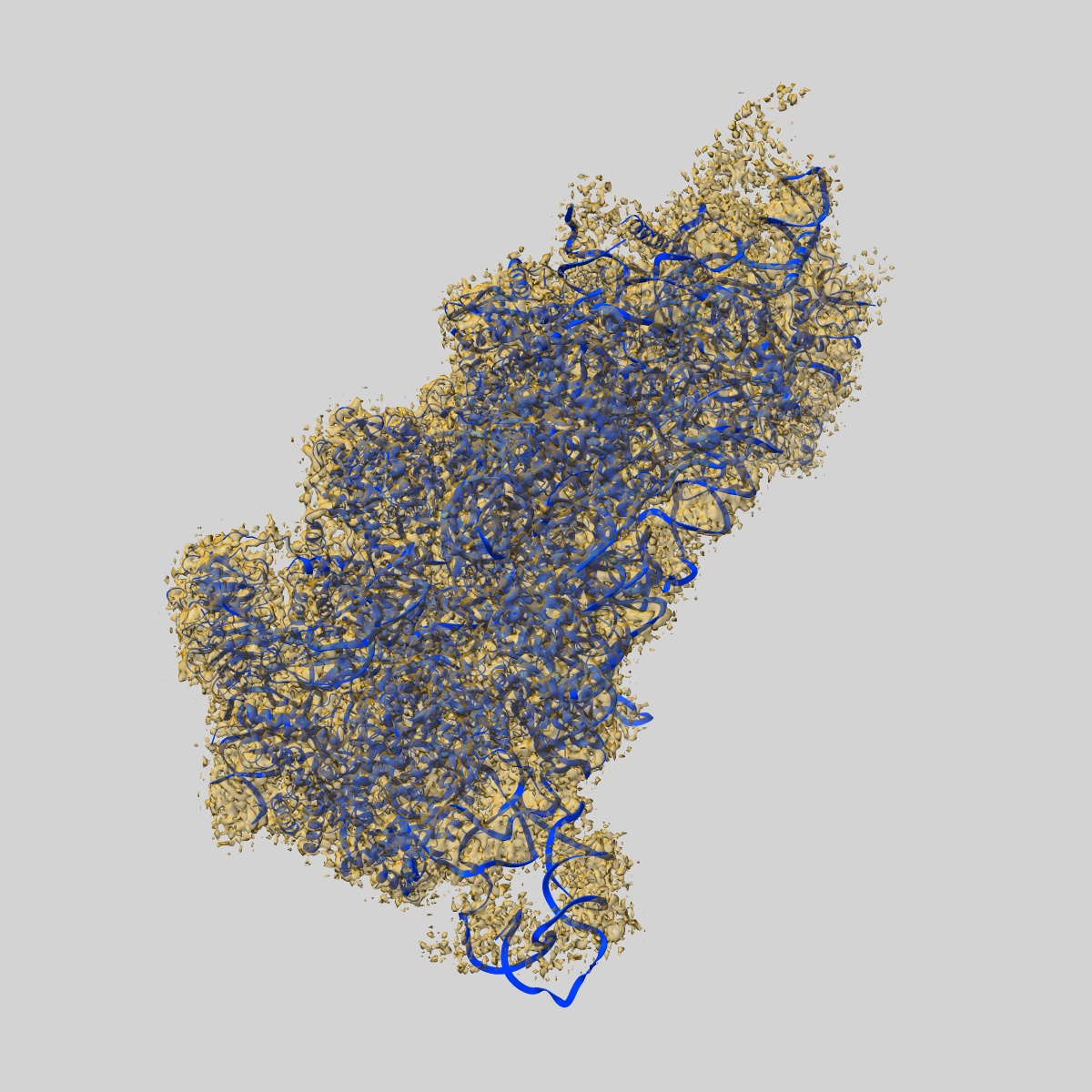
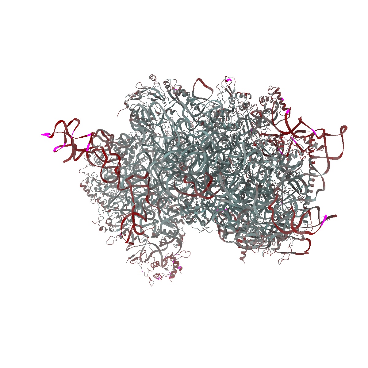
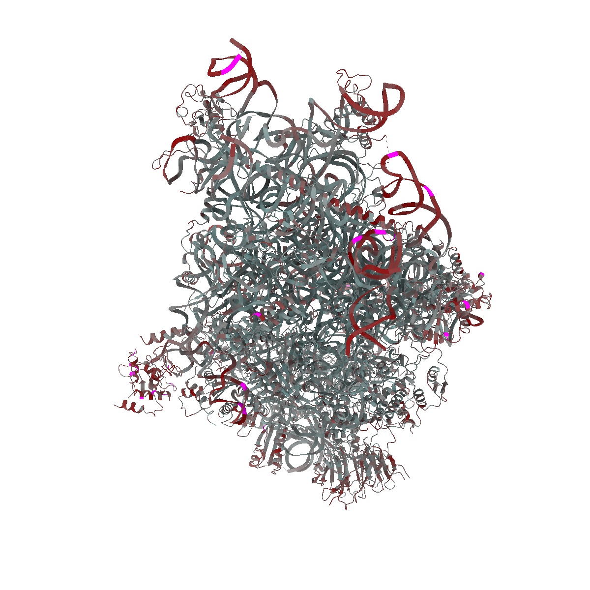
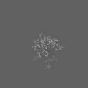
Structure of a mammalian small ribosomal subunit in complex with the Israeli Acute Paralysis Virus IRES (Class 2)
EMD-20249
Single-particle3.2 Å
 Deposition: 27/05/2019
Map released: 18/09/2019
Last modified: 23/10/2024
Sample Organism:
Oryctolagus cuniculus,
Israeli acute paralysis virus
Sample: Structure of a mammalian small ribosomal subunit in complex with the Israeli Acute Paralysis Virus IRES (Class 2)
Fitted models: 6p4h (Avg. Q-score: 0.469)
Deposition Authors: Acosta-Reyes FJ, Neupane R
Sample: Structure of a mammalian small ribosomal subunit in complex with the Israeli Acute Paralysis Virus IRES (Class 2)
Fitted models: 6p4h (Avg. Q-score: 0.469)
Deposition Authors: Acosta-Reyes FJ, Neupane R

The Israeli acute paralysis virus IRES captures host ribosomes by mimicking a ribosomal state with hybrid tRNAs.
Abstract:
Colony collapse disorder (CCD) is a multi-faceted syndrome decimating bee populations worldwide, and a group of viruses of the widely distributed Dicistroviridae family have been identified as a causing agent of CCD. This family of viruses employs non-coding RNA sequences, called internal ribosomal entry sites (IRESs), to precisely exploit the host machinery for viral protein production. Using single-particle cryo-electron microscopy (cryo-EM), we have characterized how the IRES of Israeli acute paralysis virus (IAPV) intergenic region captures and redirects translating ribosomes toward viral RNA messages. We reconstituted two in vitro reactions targeting a pre-translocation and a post-translocation state of the IAPV-IRES in the ribosome, allowing us to identify six structures using image processing classification methods. From these, we reconstructed the trajectory of IAPV-IRES from the early small subunit recruitment to the final post-translocated state in the ribosome. An early commitment of IRES/ribosome complexes for global pre-translocation mimicry explains the high efficiency observed for this IRES. Efforts directed toward fighting CCD by targeting the IAPV-IRES using RNA-interference technology are underway, and the structural framework presented here may assist in further refining these approaches.
Colony collapse disorder (CCD) is a multi-faceted syndrome decimating bee populations worldwide, and a group of viruses of the widely distributed Dicistroviridae family have been identified as a causing agent of CCD. This family of viruses employs non-coding RNA sequences, called internal ribosomal entry sites (IRESs), to precisely exploit the host machinery for viral protein production. Using single-particle cryo-electron microscopy (cryo-EM), we have characterized how the IRES of Israeli acute paralysis virus (IAPV) intergenic region captures and redirects translating ribosomes toward viral RNA messages. We reconstituted two in vitro reactions targeting a pre-translocation and a post-translocation state of the IAPV-IRES in the ribosome, allowing us to identify six structures using image processing classification methods. From these, we reconstructed the trajectory of IAPV-IRES from the early small subunit recruitment to the final post-translocated state in the ribosome. An early commitment of IRES/ribosome complexes for global pre-translocation mimicry explains the high efficiency observed for this IRES. Efforts directed toward fighting CCD by targeting the IAPV-IRES using RNA-interference technology are underway, and the structural framework presented here may assist in further refining these approaches.