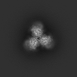
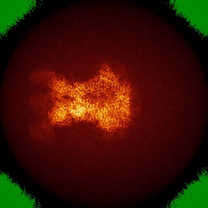
EMD-22110
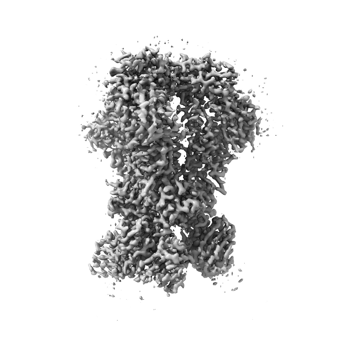
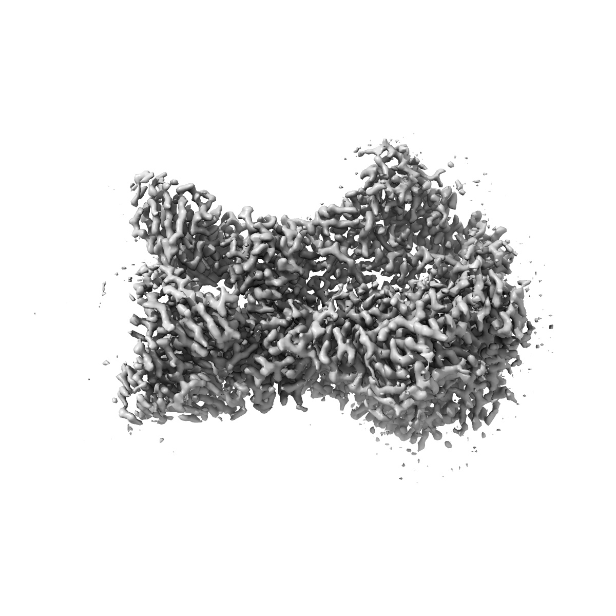
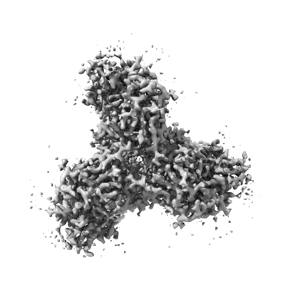
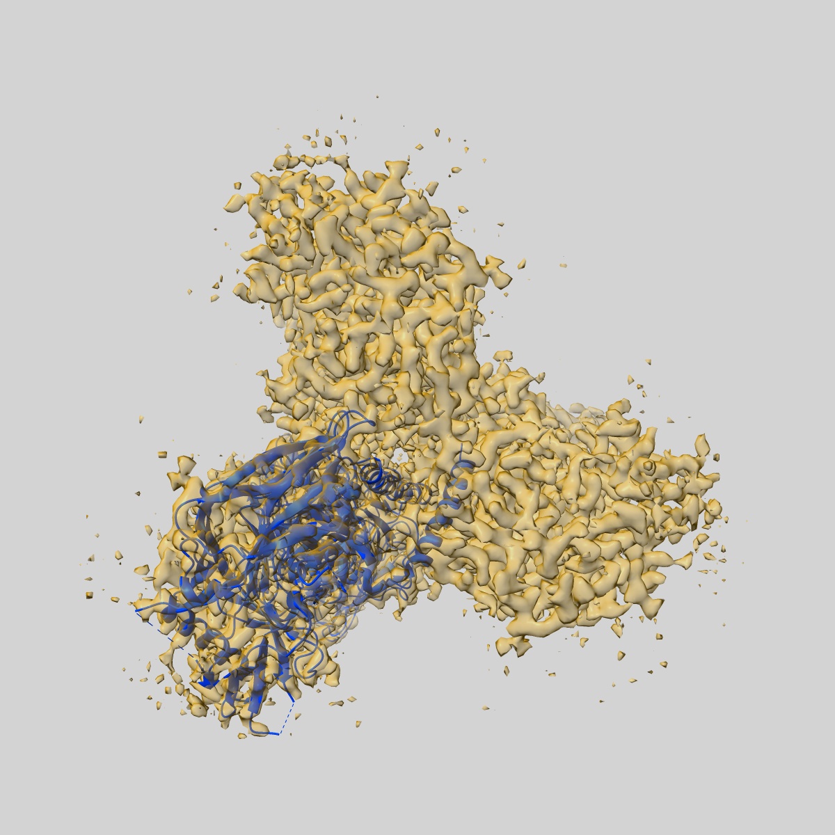
HIV-1 Envelope Glycoprotein BG505 SOSIP.664 expressed in HEK293S cells in complex with RM20A3 Fab
EMD-22110
Single-particle3.2 Å
 Deposition: 03/06/2020
Map released: 04/11/2020
Last modified: 23/10/2024
Sample Organism:
Human immunodeficiency virus 1,
Macaca mulatta
Sample: HIV-1 Envelope Glycoprotein BG505 SOSIP.664 expressed in HEK293S cells in complex with RM20A3 Fab
Fitted models: 6x9t (Avg. Q-score: 0.545)
Deposition Authors: Berndsen ZT ,
Ward AB
,
Ward AB
Sample: HIV-1 Envelope Glycoprotein BG505 SOSIP.664 expressed in HEK293S cells in complex with RM20A3 Fab
Fitted models: 6x9t (Avg. Q-score: 0.545)
Deposition Authors: Berndsen ZT
 ,
Ward AB
,
Ward AB
Visualization of the HIV-1 Env glycan shield across scales.
Berndsen ZT ,
Chakraborty S ,
Wang X ,
Cottrell CA ,
Torres JL ,
Diedrich JK ,
Lopez CA ,
Yates 3rd JR ,
van Gils MJ ,
Paulson JC ,
Gnanakaran S ,
Ward AB
(2020) PNAS , 117 , 28014 - 28025
,
Chakraborty S ,
Wang X ,
Cottrell CA ,
Torres JL ,
Diedrich JK ,
Lopez CA ,
Yates 3rd JR ,
van Gils MJ ,
Paulson JC ,
Gnanakaran S ,
Ward AB
(2020) PNAS , 117 , 28014 - 28025
Abstract:
The dense array of N-linked glycans on the HIV-1 envelope glycoprotein (Env), known as the "glycan shield," is a key determinant of immunogenicity, yet intrinsic heterogeneity confounds typical structure-function analysis. Here, we present an integrated approach of single-particle electron cryomicroscopy (cryo-EM), computational modeling, and site-specific mass spectrometry (MS) to probe glycan shield structure and behavior at multiple levels. We found that dynamics lead to an extensive network of interglycan interactions that drive the formation of higher-order structure within the glycan shield. This structure defines diffuse boundaries between buried and exposed protein surface and creates a mapping of potentially immunogenic sites on Env. Analysis of Env expressed in different cell lines revealed how cryo-EM can detect subtle changes in glycan occupancy, composition, and dynamics that impact glycan shield structure and epitope accessibility. Importantly, this identified unforeseen changes in the glycan shield of Env obtained from expression in the same cell line used for vaccine production. Finally, by capturing the enzymatic deglycosylation of Env in a time-resolved manner, we found that highly connected glycan clusters are resistant to digestion and help stabilize the prefusion trimer, suggesting the glycan shield may function beyond immune evasion.
The dense array of N-linked glycans on the HIV-1 envelope glycoprotein (Env), known as the "glycan shield," is a key determinant of immunogenicity, yet intrinsic heterogeneity confounds typical structure-function analysis. Here, we present an integrated approach of single-particle electron cryomicroscopy (cryo-EM), computational modeling, and site-specific mass spectrometry (MS) to probe glycan shield structure and behavior at multiple levels. We found that dynamics lead to an extensive network of interglycan interactions that drive the formation of higher-order structure within the glycan shield. This structure defines diffuse boundaries between buried and exposed protein surface and creates a mapping of potentially immunogenic sites on Env. Analysis of Env expressed in different cell lines revealed how cryo-EM can detect subtle changes in glycan occupancy, composition, and dynamics that impact glycan shield structure and epitope accessibility. Importantly, this identified unforeseen changes in the glycan shield of Env obtained from expression in the same cell line used for vaccine production. Finally, by capturing the enzymatic deglycosylation of Env in a time-resolved manner, we found that highly connected glycan clusters are resistant to digestion and help stabilize the prefusion trimer, suggesting the glycan shield may function beyond immune evasion.