EMD-22294
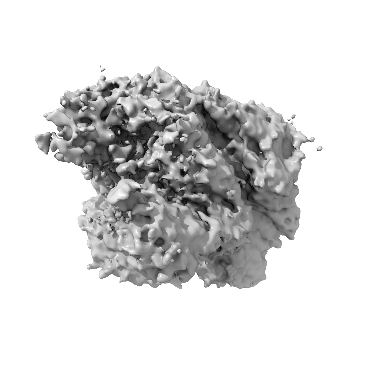
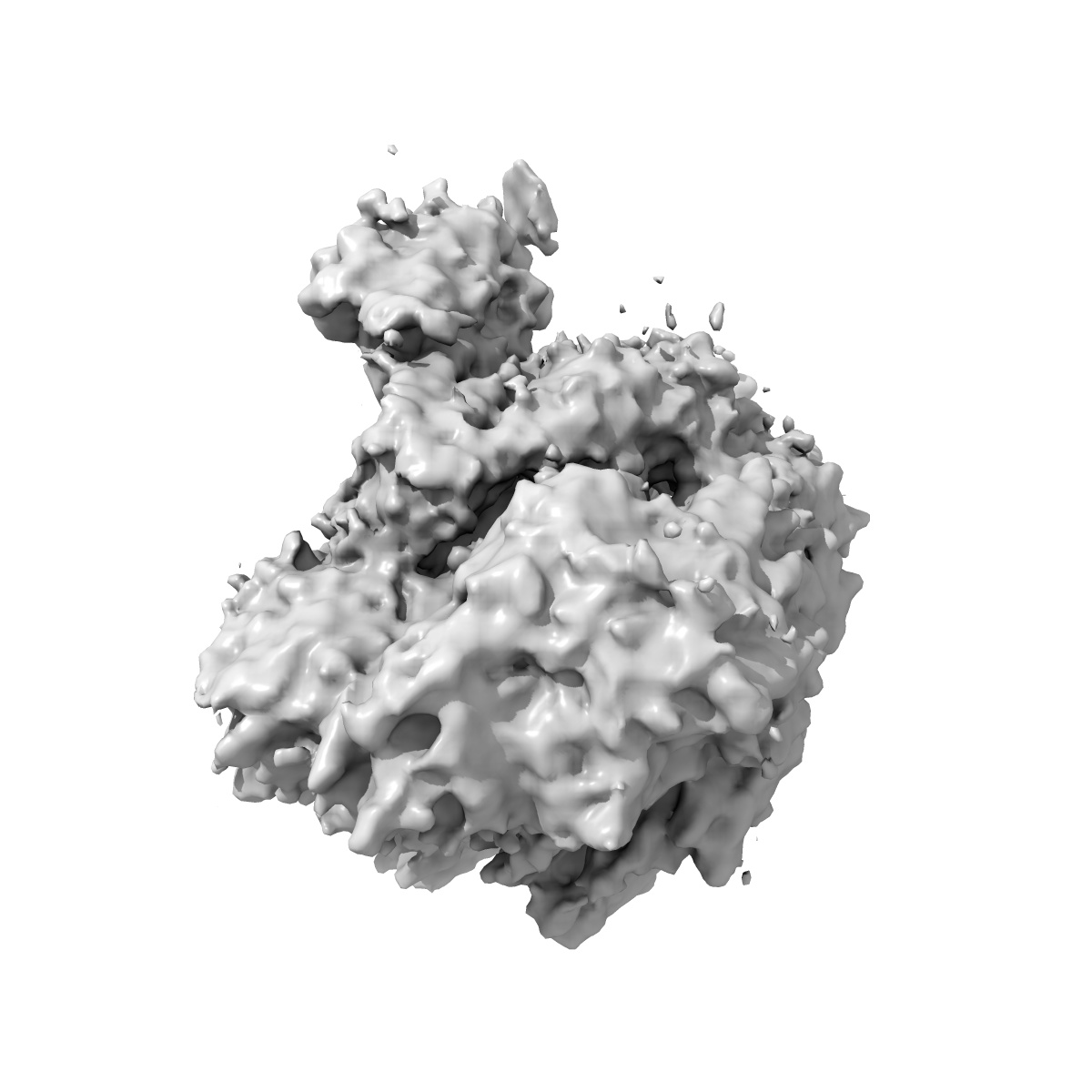
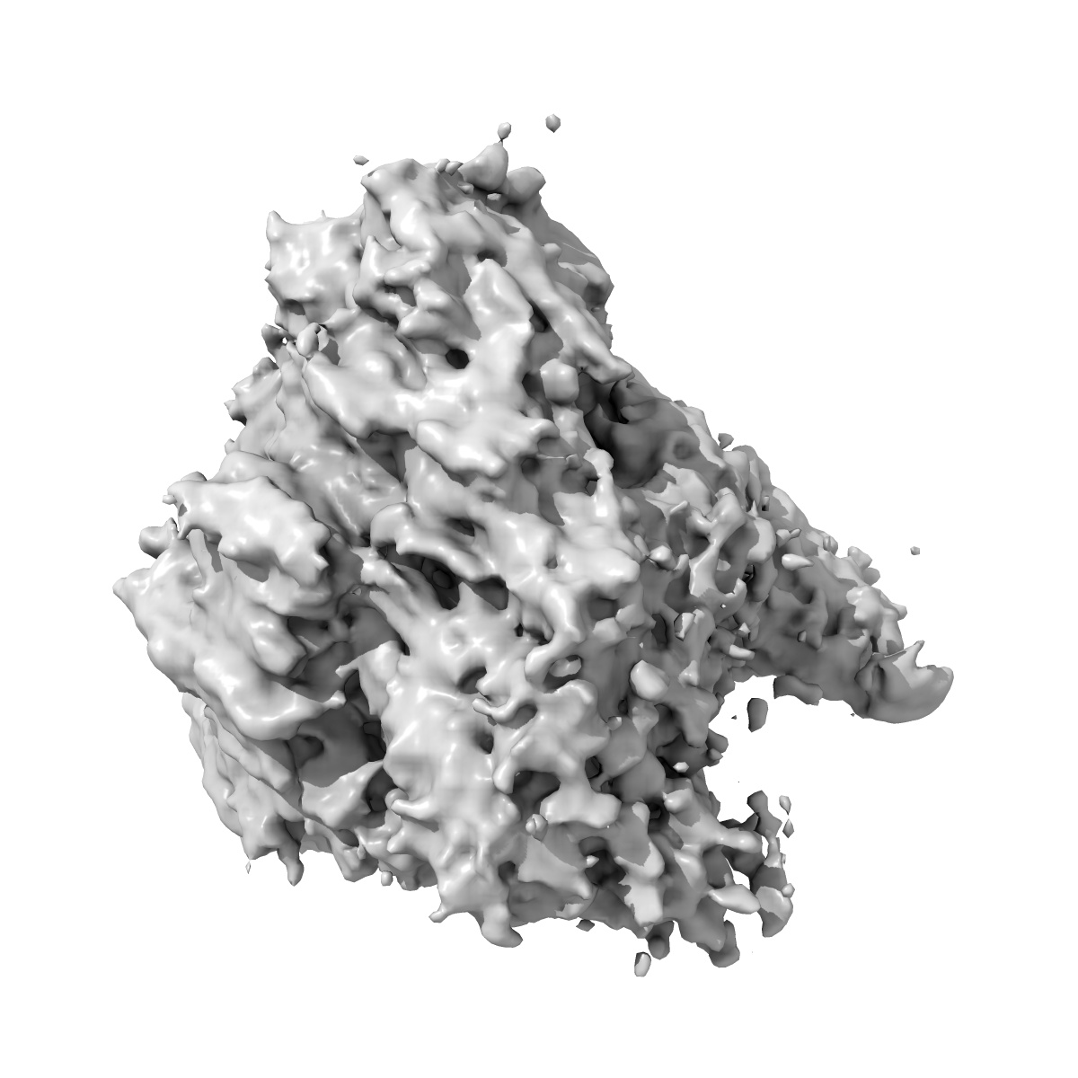
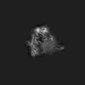
Structure of the p53/RNA polymerase II assembly
EMD-22294
Single-particle4.6 Å
 Deposition: 12/07/2020
Map released: 24/03/2021
Last modified: 06/03/2024
Sample Organism:
Homo sapiens
Sample: p53/RNA polymerase II assembly
Fitted models: 6xre (Avg. Q-score: 0.134)
Deposition Authors: Liou S-H, Singh S
Sample: p53/RNA polymerase II assembly
Fitted models: 6xre (Avg. Q-score: 0.134)
Deposition Authors: Liou S-H, Singh S
Structure of the p53/RNA polymerase II assembly.

Abstract:
The tumor suppressor p53 protein activates expression of a vast gene network in response to stress stimuli for cellular integrity. The molecular mechanism underlying how p53 targets RNA polymerase II (Pol II) to regulate transcription remains unclear. To elucidate the p53/Pol II interaction, we have determined a 4.6 Å resolution structure of the human p53/Pol II assembly via single particle cryo-electron microscopy. Our structure reveals that p53's DNA binding domain targets the upstream DNA binding site within Pol II. This association introduces conformational changes of the Pol II clamp into a further-closed state. A cavity was identified between p53 and Pol II that could possibly host DNA. The transactivation domain of p53 binds the surface of Pol II's jaw that contacts downstream DNA. These findings suggest that p53's functional domains directly regulate DNA binding activity of Pol II to mediate transcription, thereby providing insights into p53-regulated gene expression.
The tumor suppressor p53 protein activates expression of a vast gene network in response to stress stimuli for cellular integrity. The molecular mechanism underlying how p53 targets RNA polymerase II (Pol II) to regulate transcription remains unclear. To elucidate the p53/Pol II interaction, we have determined a 4.6 Å resolution structure of the human p53/Pol II assembly via single particle cryo-electron microscopy. Our structure reveals that p53's DNA binding domain targets the upstream DNA binding site within Pol II. This association introduces conformational changes of the Pol II clamp into a further-closed state. A cavity was identified between p53 and Pol II that could possibly host DNA. The transactivation domain of p53 binds the surface of Pol II's jaw that contacts downstream DNA. These findings suggest that p53's functional domains directly regulate DNA binding activity of Pol II to mediate transcription, thereby providing insights into p53-regulated gene expression.