EMD-2244
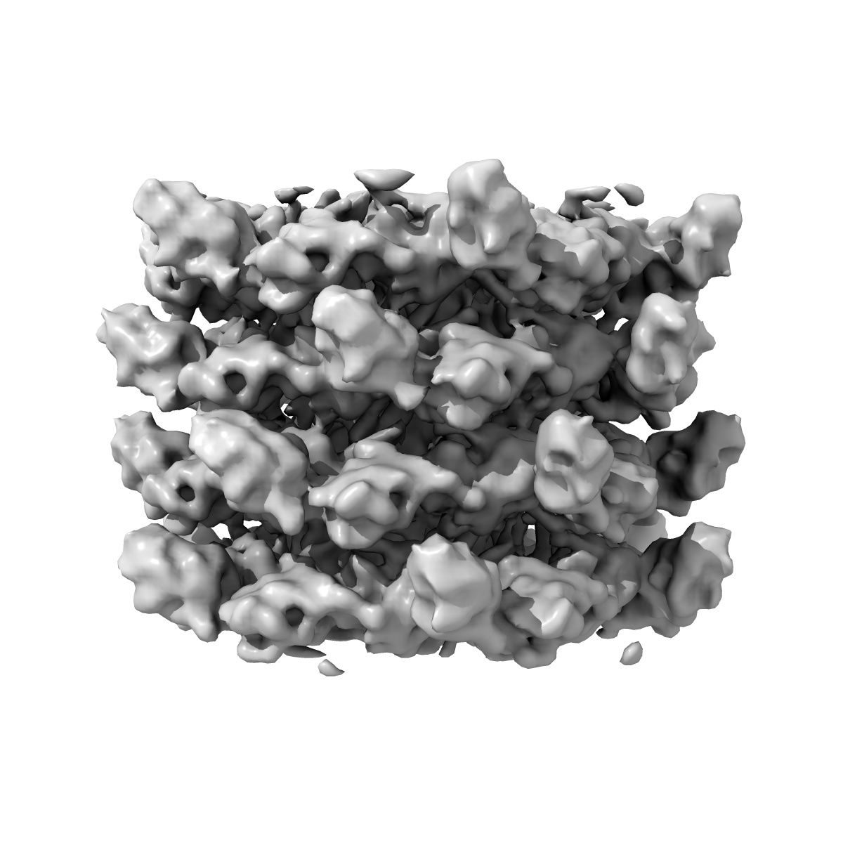
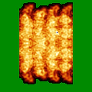
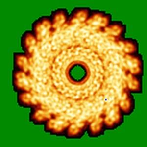
Cryo-electron microscopy of phirsl1 jumbo phage
EMD-2244
Helical reconstruction9.6 Å
 Deposition: 18/12/2012
Map released: 20/02/2013
Last modified: 20/04/2016
Sample Organism:
Ralstonia phage RSL1
Sample: phirsl1 helical portion of the tail
Deposition Authors: Effantin G, Hamasaki R, Kawasaki T, Bacia M, Moriscot C, Weissenhorn W, Yamada T, Schoehn G
Sample: phirsl1 helical portion of the tail
Deposition Authors: Effantin G, Hamasaki R, Kawasaki T, Bacia M, Moriscot C, Weissenhorn W, Yamada T, Schoehn G
Cryo-Electron Microscopy Three-Dimensional Structure of the Jumbo Phage PhiRSL1 Infecting the Phytopathogen Ralstonia solanacearum.
Effantin G,
Hamasaki R,
Kawasaki T  ,
Bacia M,
Moriscot C ,
Weissenhorn W,
Yamada T,
Schoehn G
,
Bacia M,
Moriscot C ,
Weissenhorn W,
Yamada T,
Schoehn G
(2013) Structure , 21 , 298 - 305
 ,
Bacia M,
Moriscot C ,
Weissenhorn W,
Yamada T,
Schoehn G
,
Bacia M,
Moriscot C ,
Weissenhorn W,
Yamada T,
Schoehn G
(2013) Structure , 21 , 298 - 305
Abstract:
ϕRSL1 jumbo phage belongs to a new class of viruses within the Myoviridae family. Here, we report its three-dimensional structure determined by electron cryo microscopy. The icosahedral capsid, the tail helical portion, and the complete tail appendage were reconstructed separately to resolutions of 9 Å, 9 Å, and 28 Å, respectively. The head is rather complex and formed by at least five different proteins, whereas the major capsid proteins resemble those from HK97, despite low sequence conservation. The helical tail structure demonstrates its close relationship to T4 sheath proteins and provides evidence for an evolutionary link of the inner tail tube to the bacterial type VI secretion apparatus. Long fibers extend from the collar region, and their length is consistent with reaching the host cell surface upon tail contraction. Our structural analyses indicate that ϕRSL1 is an unusual member of the Myoviridae that employs conserved protein machines related to different phages and bacteria.
ϕRSL1 jumbo phage belongs to a new class of viruses within the Myoviridae family. Here, we report its three-dimensional structure determined by electron cryo microscopy. The icosahedral capsid, the tail helical portion, and the complete tail appendage were reconstructed separately to resolutions of 9 Å, 9 Å, and 28 Å, respectively. The head is rather complex and formed by at least five different proteins, whereas the major capsid proteins resemble those from HK97, despite low sequence conservation. The helical tail structure demonstrates its close relationship to T4 sheath proteins and provides evidence for an evolutionary link of the inner tail tube to the bacterial type VI secretion apparatus. Long fibers extend from the collar region, and their length is consistent with reaching the host cell surface upon tail contraction. Our structural analyses indicate that ϕRSL1 is an unusual member of the Myoviridae that employs conserved protein machines related to different phages and bacteria.