EMD-22964
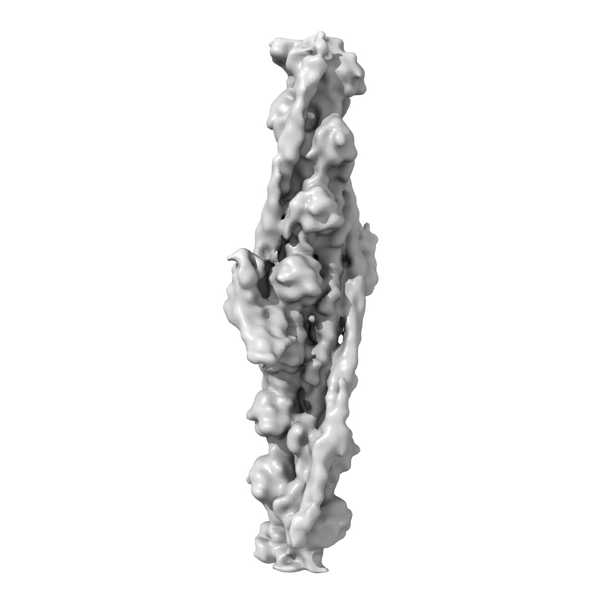
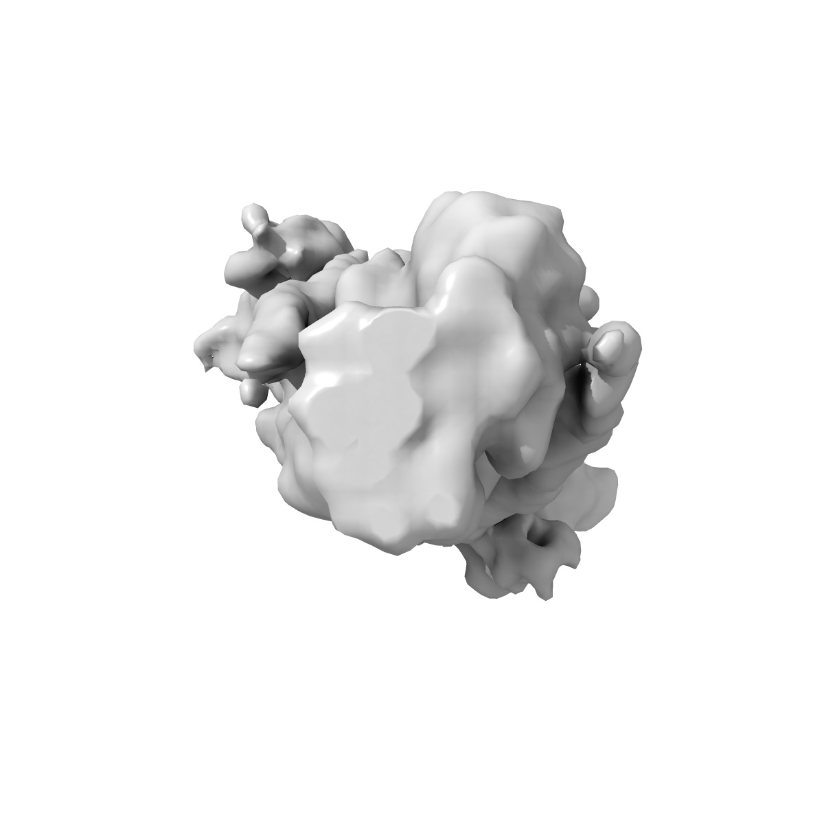
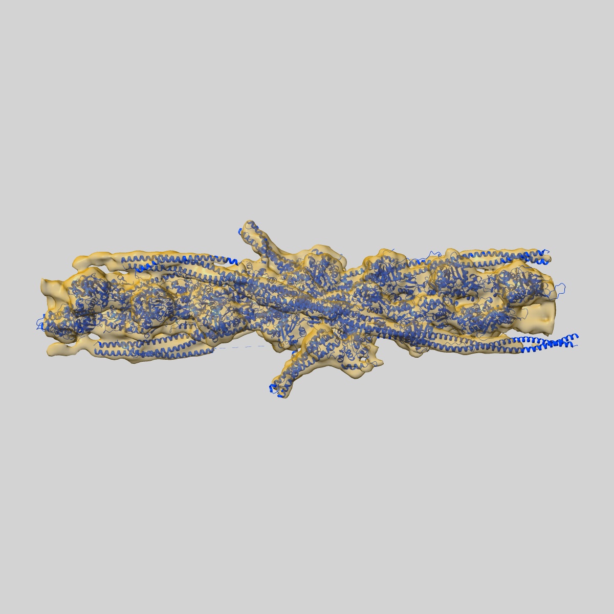
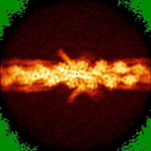
Structure of cardiac native thin filament at pCa=5.8 having upper and lower troponins in Ca2+ free state
EMD-22964
Single-particle8.0 Å
 Deposition: 06/11/2020
Map released: 24/03/2021
Last modified: 06/03/2024
Sample Organism:
Sus scrofa
Sample: cardiac native thin filament
Fitted models: 7ko4 (Avg. Q-score: 0.106)
Deposition Authors: Galkin VE ,
Risi CM
,
Risi CM
Sample: cardiac native thin filament
Fitted models: 7ko4 (Avg. Q-score: 0.106)
Deposition Authors: Galkin VE
 ,
Risi CM
,
Risi CM
The structure of the native cardiac thin filament at systolic Ca 2+ levels.
Risi CM,
Pepper I,
Belknap B,
Landim-Vieira M ,
White HD ,
Dryden K ,
Pinto JR ,
Chase PB ,
Galkin VE
(2021) PNAS , 118
,
White HD ,
Dryden K ,
Pinto JR ,
Chase PB ,
Galkin VE
(2021) PNAS , 118
Abstract:
Every heartbeat relies on cyclical interactions between myosin thick and actin thin filaments orchestrated by rising and falling Ca2+ levels. Thin filaments are comprised of two actin strands, each harboring equally separated troponin complexes, which bind Ca2+ to move tropomyosin cables away from the myosin binding sites and, thus, activate systolic contraction. Recently, structures of thin filaments obtained at low (pCa ∼9) or high (pCa ∼3) Ca2+ levels revealed the transition between the Ca2+-free and Ca2+-bound states. However, in working cardiac muscle, Ca2+ levels fluctuate at intermediate values between pCa ∼6 and pCa ∼7. The structure of the thin filament at physiological Ca2+ levels is unknown. We used cryoelectron microscopy and statistical analysis to reveal the structure of the cardiac thin filament at systolic pCa = 5.8. We show that the two strands of the thin filament consist of a mixture of regulatory units, which are composed of Ca2+-free, Ca2+-bound, or mixed (e.g., Ca2+ free on one side and Ca2+ bound on the other side) troponin complexes. We traced troponin complex conformations along and across individual thin filaments to directly determine the structural composition of the cardiac native thin filament at systolic Ca2+ levels. We demonstrate that the two thin filament strands are activated stochastically with short-range cooperativity evident only on one of the two strands. Our findings suggest a mechanism by which cardiac muscle is regulated by narrow range Ca2+ fluctuations.
Every heartbeat relies on cyclical interactions between myosin thick and actin thin filaments orchestrated by rising and falling Ca2+ levels. Thin filaments are comprised of two actin strands, each harboring equally separated troponin complexes, which bind Ca2+ to move tropomyosin cables away from the myosin binding sites and, thus, activate systolic contraction. Recently, structures of thin filaments obtained at low (pCa ∼9) or high (pCa ∼3) Ca2+ levels revealed the transition between the Ca2+-free and Ca2+-bound states. However, in working cardiac muscle, Ca2+ levels fluctuate at intermediate values between pCa ∼6 and pCa ∼7. The structure of the thin filament at physiological Ca2+ levels is unknown. We used cryoelectron microscopy and statistical analysis to reveal the structure of the cardiac thin filament at systolic pCa = 5.8. We show that the two strands of the thin filament consist of a mixture of regulatory units, which are composed of Ca2+-free, Ca2+-bound, or mixed (e.g., Ca2+ free on one side and Ca2+ bound on the other side) troponin complexes. We traced troponin complex conformations along and across individual thin filaments to directly determine the structural composition of the cardiac native thin filament at systolic Ca2+ levels. We demonstrate that the two thin filament strands are activated stochastically with short-range cooperativity evident only on one of the two strands. Our findings suggest a mechanism by which cardiac muscle is regulated by narrow range Ca2+ fluctuations.