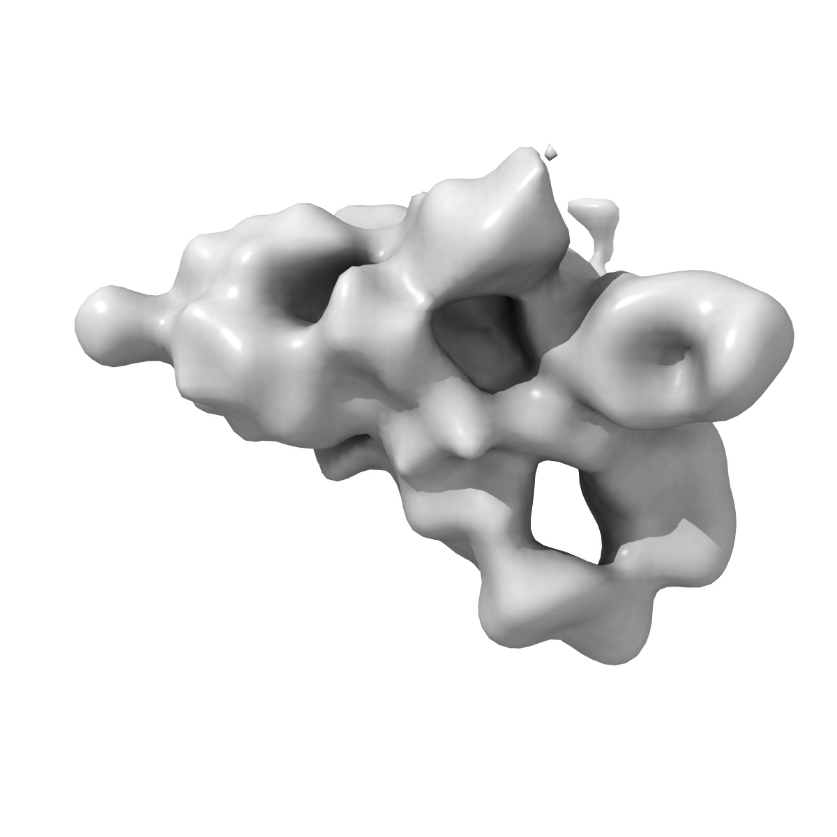
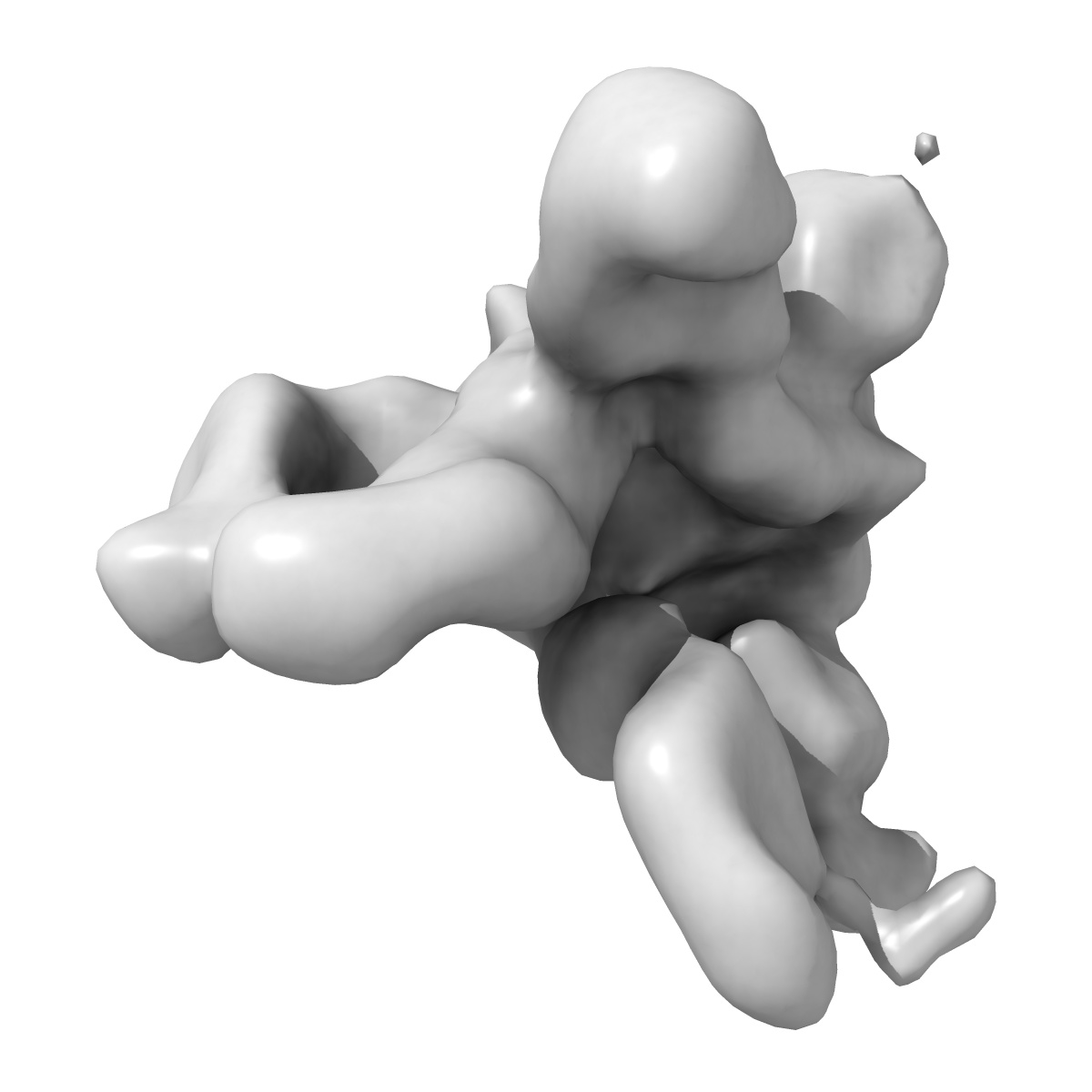
EMD-22970
Negative stain electron microscopy of 2P SARS-CoV-2 spike ectodomain in complex with Fabs DH1043 and DH1050.1
EMD-22970
Single-particle17.5 Å
 Deposition: 07/11/2020
Map released: 18/11/2020
Last modified: 01/09/2021
Sample Organism:
Homo sapiens,
Severe acute respiratory syndrome coronavirus 2
Sample: 2P SARS-CoV-2 spike ectodomain in complex with Fabs DH1043 and DH1050.1
Deposition Authors: Edwards RJ, Mansouri K
Sample: 2P SARS-CoV-2 spike ectodomain in complex with Fabs DH1043 and DH1050.1
Deposition Authors: Edwards RJ, Mansouri K
In vitro and in vivo functions of SARS-CoV-2 infection-enhancing and neutralizing antibodies.
Li D,
Edwards RJ  ,
Manne K,
Martinez DR,
Schafer A,
Alam SM,
Wiehe K,
Lu X,
Parks R,
Sutherland LL,
McDanal C,
Perez LG,
Mansouri K ,
Gobeil SMC,
Janowska K,
Stalls V,
Kopp M,
Cai F,
Lee E,
Foulger A,
Hernandez GE,
Sanzone A,
Tilahun K ,
Jiang C,
Tse LV,
Bock KW,
Minai M,
Nagata BM,
Cronin K,
Gee-Lai V,
Deyton M,
Barr M,
Von Holle T,
Macintyre AN,
Stover E,
Feldman J,
Hauser BM ,
Caradonna TM ,
Scobey TD,
Rountree W,
Wang Y,
Moody MA,
Cain DW,
DeMarco CT,
Denny TN,
Woods CW,
Petzold EW,
Schmidt AG,
Teng IT,
Zhou T,
Kwong PD,
Mascola JR,
Graham BS,
Moore IN,
Seder R,
Andersen H,
Lewis MG,
Montefiori DC,
Sempowski GD ,
Baric RS,
Acharya P,
Haynes BF,
Saunders KO
,
Manne K,
Martinez DR,
Schafer A,
Alam SM,
Wiehe K,
Lu X,
Parks R,
Sutherland LL,
McDanal C,
Perez LG,
Mansouri K ,
Gobeil SMC,
Janowska K,
Stalls V,
Kopp M,
Cai F,
Lee E,
Foulger A,
Hernandez GE,
Sanzone A,
Tilahun K ,
Jiang C,
Tse LV,
Bock KW,
Minai M,
Nagata BM,
Cronin K,
Gee-Lai V,
Deyton M,
Barr M,
Von Holle T,
Macintyre AN,
Stover E,
Feldman J,
Hauser BM ,
Caradonna TM ,
Scobey TD,
Rountree W,
Wang Y,
Moody MA,
Cain DW,
DeMarco CT,
Denny TN,
Woods CW,
Petzold EW,
Schmidt AG,
Teng IT,
Zhou T,
Kwong PD,
Mascola JR,
Graham BS,
Moore IN,
Seder R,
Andersen H,
Lewis MG,
Montefiori DC,
Sempowski GD ,
Baric RS,
Acharya P,
Haynes BF,
Saunders KO
(2021) Cell , 184 , 4203 - 4219.e32
 ,
Manne K,
Martinez DR,
Schafer A,
Alam SM,
Wiehe K,
Lu X,
Parks R,
Sutherland LL,
McDanal C,
Perez LG,
Mansouri K ,
Gobeil SMC,
Janowska K,
Stalls V,
Kopp M,
Cai F,
Lee E,
Foulger A,
Hernandez GE,
Sanzone A,
Tilahun K ,
Jiang C,
Tse LV,
Bock KW,
Minai M,
Nagata BM,
Cronin K,
Gee-Lai V,
Deyton M,
Barr M,
Von Holle T,
Macintyre AN,
Stover E,
Feldman J,
Hauser BM ,
Caradonna TM ,
Scobey TD,
Rountree W,
Wang Y,
Moody MA,
Cain DW,
DeMarco CT,
Denny TN,
Woods CW,
Petzold EW,
Schmidt AG,
Teng IT,
Zhou T,
Kwong PD,
Mascola JR,
Graham BS,
Moore IN,
Seder R,
Andersen H,
Lewis MG,
Montefiori DC,
Sempowski GD ,
Baric RS,
Acharya P,
Haynes BF,
Saunders KO
,
Manne K,
Martinez DR,
Schafer A,
Alam SM,
Wiehe K,
Lu X,
Parks R,
Sutherland LL,
McDanal C,
Perez LG,
Mansouri K ,
Gobeil SMC,
Janowska K,
Stalls V,
Kopp M,
Cai F,
Lee E,
Foulger A,
Hernandez GE,
Sanzone A,
Tilahun K ,
Jiang C,
Tse LV,
Bock KW,
Minai M,
Nagata BM,
Cronin K,
Gee-Lai V,
Deyton M,
Barr M,
Von Holle T,
Macintyre AN,
Stover E,
Feldman J,
Hauser BM ,
Caradonna TM ,
Scobey TD,
Rountree W,
Wang Y,
Moody MA,
Cain DW,
DeMarco CT,
Denny TN,
Woods CW,
Petzold EW,
Schmidt AG,
Teng IT,
Zhou T,
Kwong PD,
Mascola JR,
Graham BS,
Moore IN,
Seder R,
Andersen H,
Lewis MG,
Montefiori DC,
Sempowski GD ,
Baric RS,
Acharya P,
Haynes BF,
Saunders KO
(2021) Cell , 184 , 4203 - 4219.e32
Abstract:
SARS-CoV-2-neutralizing antibodies (NAbs) protect against COVID-19. A concern regarding SARS-CoV-2 antibodies is whether they mediate disease enhancement. Here, we isolated NAbs against the receptor-binding domain (RBD) or the N-terminal domain (NTD) of SARS-CoV-2 spike from individuals with acute or convalescent SARS-CoV-2 or a history of SARS-CoV infection. Cryo-electron microscopy of RBD and NTD antibodies demonstrated function-specific modes of binding. Select RBD NAbs also demonstrated Fc receptor-γ (FcγR)-mediated enhancement of virus infection in vitro, while five non-neutralizing NTD antibodies mediated FcγR-independent in vitro infection enhancement. However, both types of infection-enhancing antibodies protected from SARS-CoV-2 replication in monkeys and mice. Three of 46 monkeys infused with enhancing antibodies had higher lung inflammation scores compared to controls. One monkey had alveolar edema and elevated bronchoalveolar lavage inflammatory cytokines. Thus, while in vitro antibody-enhanced infection does not necessarily herald enhanced infection in vivo, increased lung inflammation can rarely occur in SARS-CoV-2 antibody-infused macaques.
SARS-CoV-2-neutralizing antibodies (NAbs) protect against COVID-19. A concern regarding SARS-CoV-2 antibodies is whether they mediate disease enhancement. Here, we isolated NAbs against the receptor-binding domain (RBD) or the N-terminal domain (NTD) of SARS-CoV-2 spike from individuals with acute or convalescent SARS-CoV-2 or a history of SARS-CoV infection. Cryo-electron microscopy of RBD and NTD antibodies demonstrated function-specific modes of binding. Select RBD NAbs also demonstrated Fc receptor-γ (FcγR)-mediated enhancement of virus infection in vitro, while five non-neutralizing NTD antibodies mediated FcγR-independent in vitro infection enhancement. However, both types of infection-enhancing antibodies protected from SARS-CoV-2 replication in monkeys and mice. Three of 46 monkeys infused with enhancing antibodies had higher lung inflammation scores compared to controls. One monkey had alveolar edema and elevated bronchoalveolar lavage inflammatory cytokines. Thus, while in vitro antibody-enhanced infection does not necessarily herald enhanced infection in vivo, increased lung inflammation can rarely occur in SARS-CoV-2 antibody-infused macaques.