EMD-24287
Cryo-ET platelet of WT mouse
EMD-24287
Tomography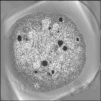 Deposition: 23/06/2021
Map released: 23/02/2022
Last modified: 23/02/2022
Using Cryo-ET to distinguish platelets during pre-acute myeloid leukemia from steady state hematopoiesis.
Wang Y,
Huo T,
Tseng YJ  ,
Dang L,
Yu Z,
Yu W,
Foulks Z,
Murdaugh RL,
Ludtke SJ ,
Nakada D ,
Wang Z
,
Dang L,
Yu Z,
Yu W,
Foulks Z,
Murdaugh RL,
Ludtke SJ ,
Nakada D ,
Wang Z
(2022) Commun Biol , 5 , 72 - 72
 ,
Dang L,
Yu Z,
Yu W,
Foulks Z,
Murdaugh RL,
Ludtke SJ ,
Nakada D ,
Wang Z
,
Dang L,
Yu Z,
Yu W,
Foulks Z,
Murdaugh RL,
Ludtke SJ ,
Nakada D ,
Wang Z
(2022) Commun Biol , 5 , 72 - 72
Abstract:
Early diagnosis of acute myeloid leukemia (AML) in the pre-leukemic stage remains a clinical challenge, as pre-leukemic patients show no symptoms, lacking any known morphological or numerical abnormalities in blood cells. Here, we demonstrate that platelets with structurally abnormal mitochondria emerge at the pre-leukemic phase of AML, preceding detectable changes in blood cell counts or detection of leukemic blasts in blood. We visualized frozen-hydrated platelets from mice at different time points during AML development in situ using electron cryo-tomography (cryo-ET) and identified intracellular organelles through an unbiased semi-automatic process followed by quantitative measurement. A large proportion of platelets exhibited changes in the overall shape and depletion of organelles in AML. Notably, 23% of platelets in pre-leukemic cells exhibit abnormal, round mitochondria with unfolded cristae, accompanied by a significant drop in ATP levels and altered expression of metabolism-related gene signatures. Our study demonstrates that detectable structural changes in pre-leukemic platelets may serve as a biomarker for the early diagnosis of AML.
Early diagnosis of acute myeloid leukemia (AML) in the pre-leukemic stage remains a clinical challenge, as pre-leukemic patients show no symptoms, lacking any known morphological or numerical abnormalities in blood cells. Here, we demonstrate that platelets with structurally abnormal mitochondria emerge at the pre-leukemic phase of AML, preceding detectable changes in blood cell counts or detection of leukemic blasts in blood. We visualized frozen-hydrated platelets from mice at different time points during AML development in situ using electron cryo-tomography (cryo-ET) and identified intracellular organelles through an unbiased semi-automatic process followed by quantitative measurement. A large proportion of platelets exhibited changes in the overall shape and depletion of organelles in AML. Notably, 23% of platelets in pre-leukemic cells exhibit abnormal, round mitochondria with unfolded cristae, accompanied by a significant drop in ATP levels and altered expression of metabolism-related gene signatures. Our study demonstrates that detectable structural changes in pre-leukemic platelets may serve as a biomarker for the early diagnosis of AML.