EMD-24587
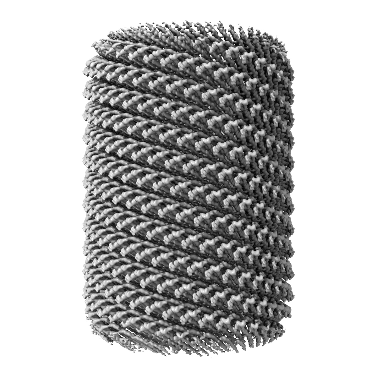
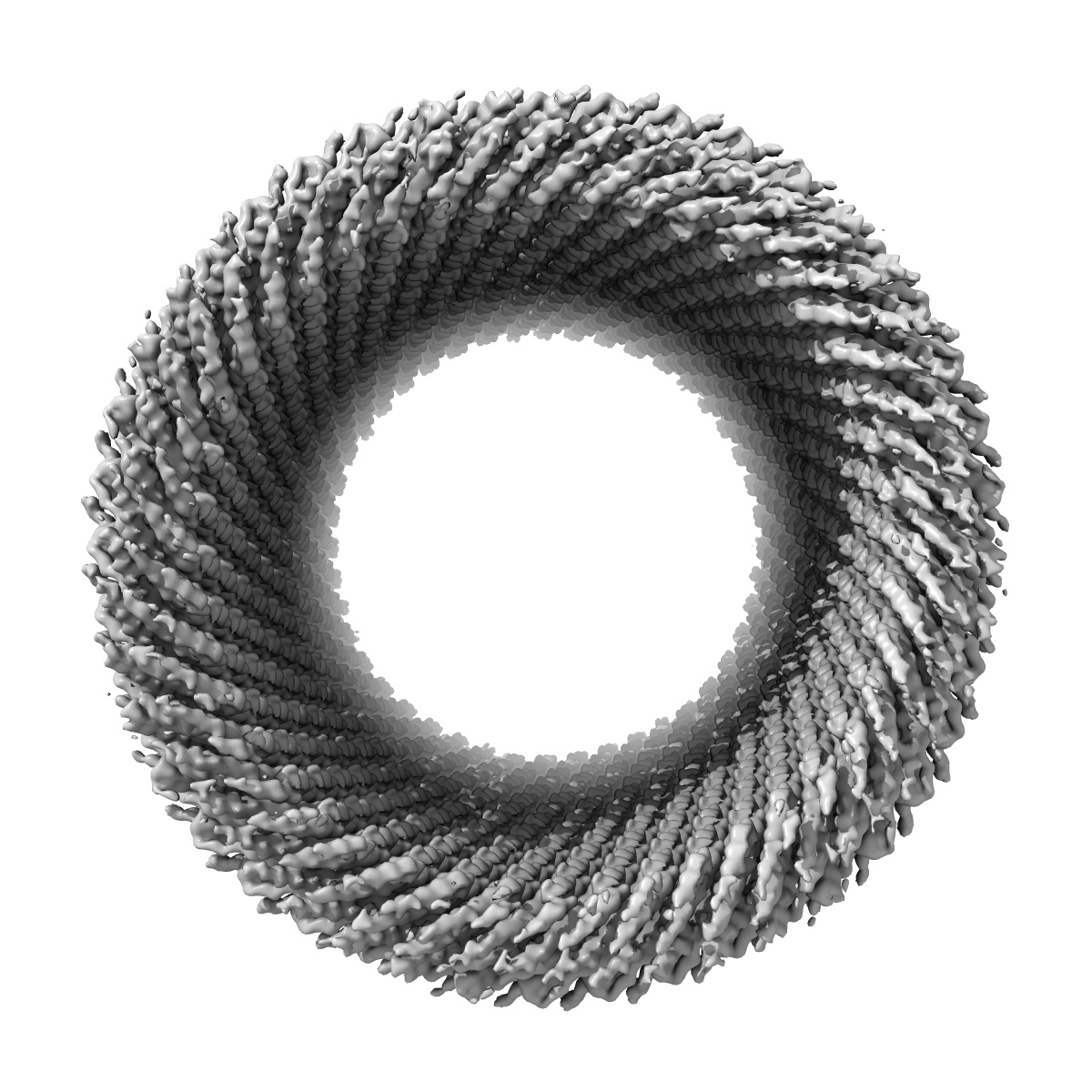
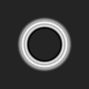
Cryo-EM reconstruction of Sulfolobus monocaudavirus SMV1, symmetry 3
EMD-24587
Helical reconstruction4.3 Å
 Deposition: 30/07/2021
Map released: 30/03/2022
Last modified: 05/06/2024
Sample Organism:
Sulfolobus monocaudavirus SMV1
Sample: Sulfolobus monocaudavirus SMV1
Fitted models: 7ro4 (Avg. Q-score: 0.378)
Deposition Authors: Wang F ,
Cvirkaite-Krupovic V
,
Cvirkaite-Krupovic V
Sample: Sulfolobus monocaudavirus SMV1
Fitted models: 7ro4 (Avg. Q-score: 0.378)
Deposition Authors: Wang F
 ,
Cvirkaite-Krupovic V
,
Cvirkaite-Krupovic V
Spindle-shaped archaeal viruses evolved from rod-shaped ancestors to package a larger genome.
Wang F ,
Cvirkaite-Krupovic V ,
Vos M,
Beltran LC,
Kreutzberger MAB,
Winter JM ,
Su Z,
Liu J,
Schouten S,
Krupovic M ,
Egelman EH
(2022) Cell , 185 , 1297
,
Cvirkaite-Krupovic V ,
Vos M,
Beltran LC,
Kreutzberger MAB,
Winter JM ,
Su Z,
Liu J,
Schouten S,
Krupovic M ,
Egelman EH
(2022) Cell , 185 , 1297
Abstract:
Spindle- or lemon-shaped viruses infect archaea in diverse environments. Due to the highly pleomorphic nature of these virions, which can be found with cylindrical tails emanating from the spindle-shaped body, structural studies of these capsids have been challenging. We have determined the atomic structure of the capsid of Sulfolobus monocaudavirus 1, a virus that infects hosts living in nearly boiling acid. A highly hydrophobic protein, likely integrated into the host membrane before the virions assemble, forms 7 strands that slide past each other in both the tails and the spindle body. We observe the discrete steps that occur as the tail tubes expand, and these are due to highly conserved quasiequivalent interactions with neighboring subunits maintained despite significant diameter changes. Our results show how helical assemblies can vary their diameters, becoming nearly spherical to package a larger genome and suggest how all spindle-shaped viruses have evolved from archaeal rod-like viruses.
Spindle- or lemon-shaped viruses infect archaea in diverse environments. Due to the highly pleomorphic nature of these virions, which can be found with cylindrical tails emanating from the spindle-shaped body, structural studies of these capsids have been challenging. We have determined the atomic structure of the capsid of Sulfolobus monocaudavirus 1, a virus that infects hosts living in nearly boiling acid. A highly hydrophobic protein, likely integrated into the host membrane before the virions assemble, forms 7 strands that slide past each other in both the tails and the spindle body. We observe the discrete steps that occur as the tail tubes expand, and these are due to highly conserved quasiequivalent interactions with neighboring subunits maintained despite significant diameter changes. Our results show how helical assemblies can vary their diameters, becoming nearly spherical to package a larger genome and suggest how all spindle-shaped viruses have evolved from archaeal rod-like viruses.