EMD-25022
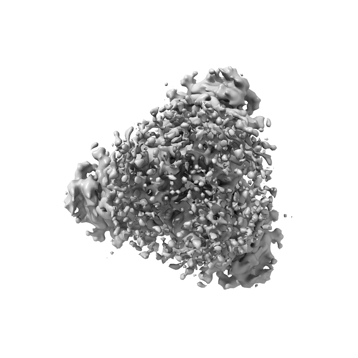
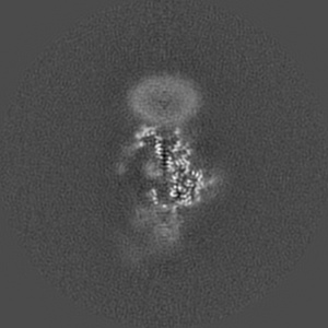
Cytoplasmic tail deleted HIV Env trimer in nanodisc
EMD-25022
Single-particle3.88 Å
 Deposition: 27/09/2021
Map released: 09/11/2022
Last modified: 20/11/2024
Sample Organism:
HIV whole-genome vector AA1305#18
Sample: HIV-1 envelope glycoprotein Env trimer
Fitted models: 7sc5 (Avg. Q-score: 0.394)
Deposition Authors: Yang S ,
Walz T
,
Walz T
Sample: HIV-1 envelope glycoprotein Env trimer
Fitted models: 7sc5 (Avg. Q-score: 0.394)
Deposition Authors: Yang S
 ,
Walz T
,
Walz T
Dynamic HIV-1 spike motion creates vulnerability for its membrane-bound tripod to antibody attack.
Yang S ,
Hiotis G,
Wang Y,
Chen J,
Wang JH,
Kim M,
Reinherz EL ,
Walz T
(2022) Nat Commun , 13 , 6393 - 6393
,
Hiotis G,
Wang Y,
Chen J,
Wang JH,
Kim M,
Reinherz EL ,
Walz T
(2022) Nat Commun , 13 , 6393 - 6393
Abstract:
Vaccines targeting HIV-1's gp160 spike protein are stymied by high viral mutation rates and structural chicanery. gp160's membrane-proximal external region (MPER) is the target of naturally arising broadly neutralizing antibodies (bnAbs), yet MPER-based vaccines fail to generate bnAbs. Here, nanodisc-embedded spike protein was investigated by cryo-electron microscopy and molecular-dynamics simulations, revealing spontaneous ectodomain tilting that creates vulnerability for HIV-1. While each MPER protomer radiates centrally towards the three-fold axis contributing to a membrane-associated tripod structure that is occluded in the upright spike, tilting provides access to the opposing MPER. Structures of spike proteins with bound 4E10 bnAb Fabs reveal that the antibody binds exposed MPER, thereby altering MPER dynamics, modifying average ectodomain tilt, and imposing strain on the viral membrane and the spike's transmembrane segments, resulting in the abrogation of membrane fusion and informing future vaccine development.
Vaccines targeting HIV-1's gp160 spike protein are stymied by high viral mutation rates and structural chicanery. gp160's membrane-proximal external region (MPER) is the target of naturally arising broadly neutralizing antibodies (bnAbs), yet MPER-based vaccines fail to generate bnAbs. Here, nanodisc-embedded spike protein was investigated by cryo-electron microscopy and molecular-dynamics simulations, revealing spontaneous ectodomain tilting that creates vulnerability for HIV-1. While each MPER protomer radiates centrally towards the three-fold axis contributing to a membrane-associated tripod structure that is occluded in the upright spike, tilting provides access to the opposing MPER. Structures of spike proteins with bound 4E10 bnAb Fabs reveal that the antibody binds exposed MPER, thereby altering MPER dynamics, modifying average ectodomain tilt, and imposing strain on the viral membrane and the spike's transmembrane segments, resulting in the abrogation of membrane fusion and informing future vaccine development.