EMD-25485
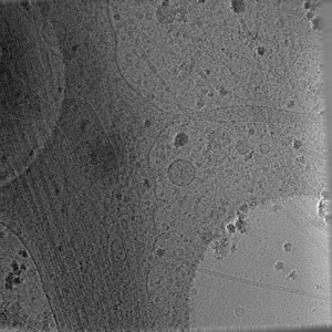
In situ cryo-electron tomography reveals local cellular machineries for axon branch development (premature branch)
EMD-25485
Tomography Deposition: 21/11/2021
Map released: 09/03/2022
Last modified: 17/01/2024
Sample Organism:
Mus musculus
Sample: Primary hippocampal neuron from mouse
Raw data: EMPIAR-10922, EMPIAR-10923
Deposition Authors: Nedozralova H ,
Basnet N ,
Ibiricu I,
Bodakuntla S ,
Biertumpfel C ,
Mizuno N
,
Basnet N ,
Ibiricu I,
Bodakuntla S ,
Biertumpfel C ,
Mizuno N
Sample: Primary hippocampal neuron from mouse
Raw data: EMPIAR-10922, EMPIAR-10923
Deposition Authors: Nedozralova H
 ,
Basnet N ,
Ibiricu I,
Bodakuntla S ,
Biertumpfel C ,
Mizuno N
,
Basnet N ,
Ibiricu I,
Bodakuntla S ,
Biertumpfel C ,
Mizuno N
In situ cryo-electron tomography reveals local cellular machineries for axon branch development.
Nedozralova H ,
Basnet N ,
Ibiricu I,
Bodakuntla S ,
Biertumpfel C ,
Mizuno N
(2022) J Cell Biol , 221
,
Basnet N ,
Ibiricu I,
Bodakuntla S ,
Biertumpfel C ,
Mizuno N
(2022) J Cell Biol , 221
Abstract:
Neurons are highly polarized cells forming an intricate network of dendrites and axons. They are shaped by the dynamic reorganization of cytoskeleton components and cellular organelles. Axon branching allows the formation of new paths and increases circuit complexity. However, our understanding of branch formation is sparse due to the lack of direct in-depth observations. Using in situ cellular cryo-electron tomography on primary mouse neurons, we directly visualized the remodeling of organelles and cytoskeleton structures at axon branches. Strikingly, branched areas functioned as hotspots concentrating organelles to support dynamic activities. Unaligned actin filaments assembled at the base of premature branches accompanied by filopodia-like protrusions. Microtubules and ER comigrated into preformed branches to support outgrowth together with accumulating compact, ∼500-nm mitochondria and locally clustered ribosomes. We obtained a roadmap of events supporting the hypothesis of local protein synthesis selectively taking place at axon branches, allowing them to serve as unique control hubs for axon development and downstream neural network formation.
Neurons are highly polarized cells forming an intricate network of dendrites and axons. They are shaped by the dynamic reorganization of cytoskeleton components and cellular organelles. Axon branching allows the formation of new paths and increases circuit complexity. However, our understanding of branch formation is sparse due to the lack of direct in-depth observations. Using in situ cellular cryo-electron tomography on primary mouse neurons, we directly visualized the remodeling of organelles and cytoskeleton structures at axon branches. Strikingly, branched areas functioned as hotspots concentrating organelles to support dynamic activities. Unaligned actin filaments assembled at the base of premature branches accompanied by filopodia-like protrusions. Microtubules and ER comigrated into preformed branches to support outgrowth together with accumulating compact, ∼500-nm mitochondria and locally clustered ribosomes. We obtained a roadmap of events supporting the hypothesis of local protein synthesis selectively taking place at axon branches, allowing them to serve as unique control hubs for axon development and downstream neural network formation.