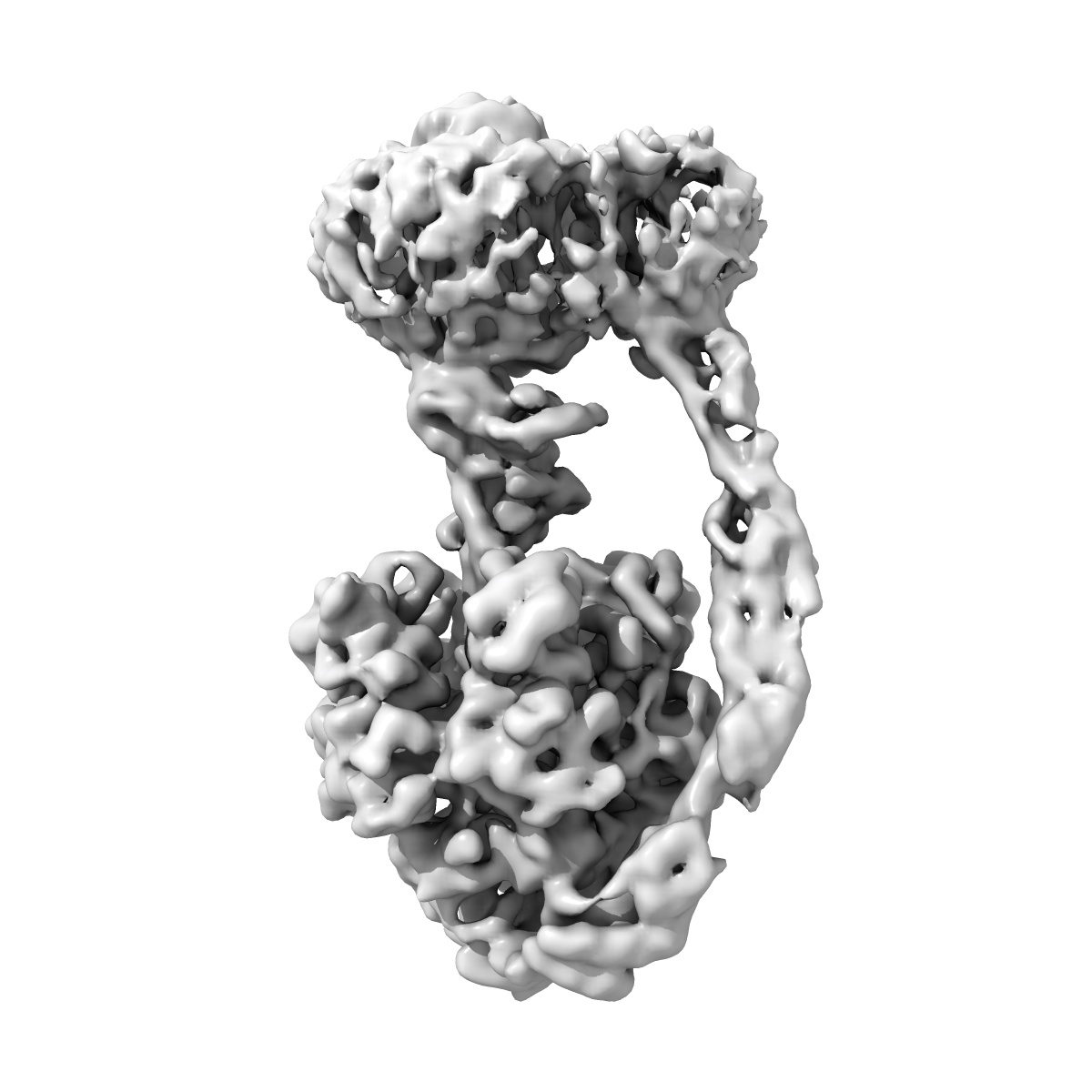
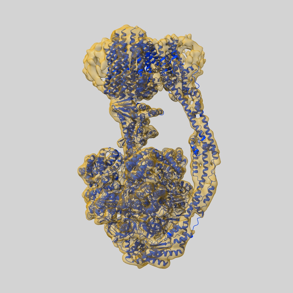
EMD-25964
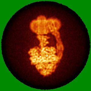
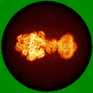
Yeast ATP synthase State 1catalytic(g) with 10 mM ATP backbone model
EMD-25964
Single-particle5.8 Å
 Deposition: 17/01/2022
Map released: 20/04/2022
Last modified: 21/02/2024
Sample Organism:
Saccharomyces cerevisiae
Sample: Yeast ATP synthase State 1catalytic(g) with 10 mM ATP backbone model
Fitted models: 7tkc (Avg. Q-score: 0.301)
Deposition Authors: Guo H ,
Rubinstein JL
,
Rubinstein JL
Sample: Yeast ATP synthase State 1catalytic(g) with 10 mM ATP backbone model
Fitted models: 7tkc (Avg. Q-score: 0.301)
Deposition Authors: Guo H
 ,
Rubinstein JL
,
Rubinstein JL
Structure of ATP synthase under strain during catalysis.
Abstract:
ATP synthases are macromolecular machines consisting of an ATP-hydrolysis-driven F1 motor and a proton-translocation-driven FO motor. The F1 and FO motors oppose each other's action on a shared rotor subcomplex and are held stationary relative to each other by a peripheral stalk. Structures of resting mitochondrial ATP synthases revealed a left-handed curvature of the peripheral stalk even though rotation of the rotor, driven by either ATP hydrolysis in F1 or proton translocation through FO, would apply a right-handed bending force to the stalk. We used cryoEM to image yeast mitochondrial ATP synthase under strain during ATP-hydrolysis-driven rotary catalysis, revealing a large deformation of the peripheral stalk. The structures show how the peripheral stalk opposes the bending force and suggests that during ATP synthesis proton translocation causes accumulation of strain in the stalk, which relaxes by driving the relative rotation of the rotor through six sub-steps within F1, leading to catalysis.
ATP synthases are macromolecular machines consisting of an ATP-hydrolysis-driven F1 motor and a proton-translocation-driven FO motor. The F1 and FO motors oppose each other's action on a shared rotor subcomplex and are held stationary relative to each other by a peripheral stalk. Structures of resting mitochondrial ATP synthases revealed a left-handed curvature of the peripheral stalk even though rotation of the rotor, driven by either ATP hydrolysis in F1 or proton translocation through FO, would apply a right-handed bending force to the stalk. We used cryoEM to image yeast mitochondrial ATP synthase under strain during ATP-hydrolysis-driven rotary catalysis, revealing a large deformation of the peripheral stalk. The structures show how the peripheral stalk opposes the bending force and suggests that during ATP synthesis proton translocation causes accumulation of strain in the stalk, which relaxes by driving the relative rotation of the rotor through six sub-steps within F1, leading to catalysis.